. 2021 Jun 19;12(1):353.
doi: 10.1186/s13287-021-02434-2.

Enhanced HSC-like cell generation from mouse pluripotent stem cells in a 3D induction system cocultured with stromal cells

Affiliations

Enhanced HSC-like cell generation from mouse pluripotent stem cells in a 3D induction system cocultured with stromal cells

Wei Shan et al. Stem Cell Res Ther. .

Abstract

Background: Decades of efforts have attempted to differentiate the pluripotent stem cells (PSCs) into truly functional hematopoietic stem cells (HSCs), yet the problems of low differentiation efficiency in vitro and poor hematopoiesis reconstitution in vivo still exist, mainly attributing to the lack of solid, reproduced, or pursued differentiation system.

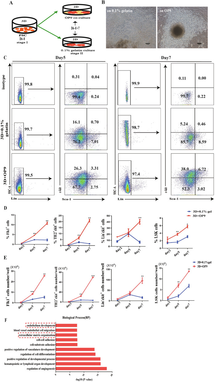
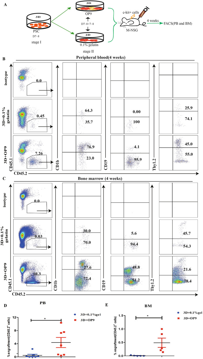
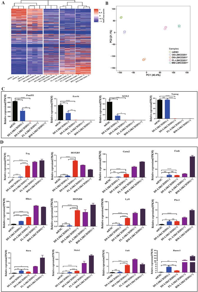
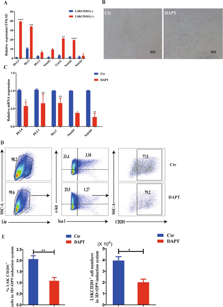
Methods: In this study, we established an in vitro differentiation system yielding in vivo hematopoietic reconstitution hematopoietic cells from mouse PSCs through a 3D induction system followed by coculture with OP9 stromal cells. The in vivo hematopoietic reconstitution potential of c-kit+ cells derived from the mouse PSCs was evaluated via m-NSG transplantation assay. Flow cytometry analysis, RNA-seq, and cell cycle analysis were used to detect the in vitro hematopoietic ability of endothelial protein C receptor (EPCR, CD201) cells generated in our induction system.

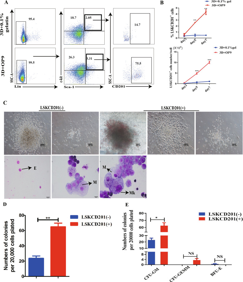
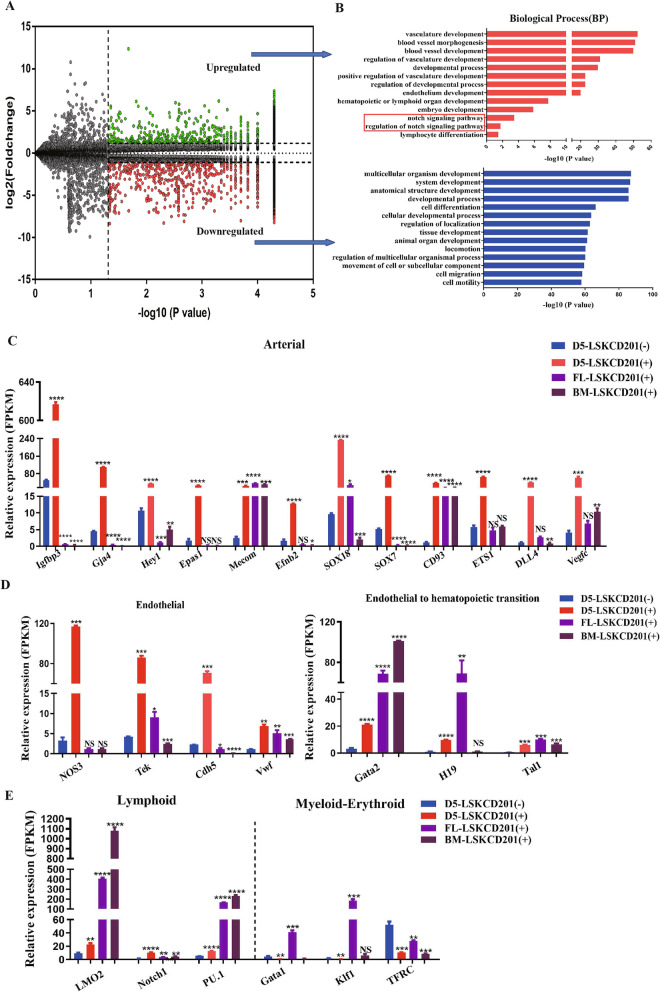
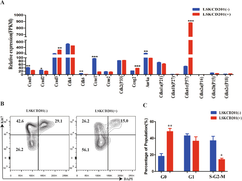
Results: The c-kit+ cells from 3D self-assembling peptide induction system followed by the OP9 coculture system possessed apparently superiority in terms of in vivo repopulating activity than that of 3D induction system followed by the 0.1% gelatin culture. We interestingly found that our 3D+OP9 system enriched a higher percentage of CD201+c-kit+cells that showed more similar HSC-like features such as transcriptome level and CFU formation ability than CD201-c-kit+cells, which have not been reported in the field of mouse PSCs hematopoietic differentiation. Moreover, CD201+ hematopoietic cells remained in a relatively slow cycling state, consistent with high expression levels of P57 and Ccng2. Further, we innovatively demonstrated that notch signaling pathway is responsible for in vitro CD201+ hematopoietic cell induction from mouse PSCs.

Conclusions: Altogether, our findings lay a foundation for improving the efficiency of hematopoietic differentiation and generating in vivo functional HSC-like cells from mouse PSCs for clinical application.

Keywords: 3D system; CD201; Hematopoiesis; Notch; Pluripotent stem cells.


Conflict of interest statement

The authors declare that they have no competing interests.

Figures

Fig. 1
Fig. 1
The 3D self-assembling peptide induction system followed by the OP9 coculture system promotes hematopoietic differentiation of mouse PSCs. A Schematic overview of mouse PSCs hematopoietic differentiation in the 3D self-assembling peptide induction system followed by the OP9 coculture system. B Representative morphology of hematopoietic cells from hematopoietic-like colonies derived from EBs in a 3D induction system followed by coculture with OP9 stromal cells and 0.1% gelatin. C Representative flow cytometry analysis of the percentage of LinSca-1+c-kit+(LSK) on day 5 and day 7 respectively (n = 3). D Statistical analysis of percentage of mesoderm cells (Flk1+), hemogenic endothelium (TIE2+c-kit+), Linc-kit+ and LSK on day 2, day 5, and day 7 respectively (n = 3). E Absolute numbers of Flk1+, TIE2+c-kit+, Linc-kit+, and LSK cells in between 3D+OP9 and 3D+0.1% gelatin on day 2, day 5, and day 7 respectively (n = 3). F Representative statistically enriched pathways of the upregulated genes in 3D+OP9 group compared with 3D+0.1% gelatin group. Data are the means ± SD from three independent experiments. Error bars represent mean ± SD of samples from three independent experiments (n = 3). NS, not significant; *p < 0.05, **p < 0.01, ***p < 0.001
Fig. 2
Fig. 2
In vivo transplanted potential of 3D+OP9 and 3D+0.1% gelatin induction system-derived c-kit+ hematopoietic cells. A Schematic representation of the transplantation strategy. BE Representative flow cytometric plots for CD45.1 and CD45.2 expression in the PB and BM from m-NSG recipient mice (CD45.1), meanwhile representative flow cytometric plots for expression of CD11b, CD19, and thy1.2 in gated CD45.2+ cells. FG Percentage CD45.2+ chimerism in the PB (left) and BM (right) from m-NSG. Data are the means ± SD from three independent experiments. Error bars represent mean ± SD of samples from at least three independent experiments (n>=3). NS, not significant; *p < 0.05
Fig. 3
Fig. 3
CD201 expression marks a cluster of functional HSC/HSPC in our 3D self-assembling peptide-mediated OP9 coculture hematopoietic induction system. A FACS analysis of the percentage of LSK and LSKCD201+ cells derived from 3D+OP9 and 3D+0.1% gelatin group on day 5. B Statistical analysis of percentage and absolute numbers LSKCD201+ cells of the 3D+OP9 and 3D+0.1% gelatin co-culture system on days 2, 5, and 7 respectively (n = 3). C Sorted Linc-kit+CD201+ cells were analyzed by a CFU assay. Representative field images of the different types of colonies obtained from Linc-kit+CD201- and Linc-kit+CD201+ cells and cellular morphology of colonies by May-Giemsa staining. Arrowheads depict indicated morphology. E, erythrocyte; M, macrophage; Mk, megakaryocyte. Scale bar =100μm. D Linc-kit+CD201+ cells formed a significantly higher number of colonies than Linc-kit+CD201- cells. E Statistical analysis of the variety of colonies between Linc-kit+CD201- cells and Linc-kit+CD201+ cells. Error bars represent mean ± SD of samples from at least three independent experiments (n = 3). NS, not significant; *p < 0.05, **p < 0.01
Fig. 4
Fig. 4
Global gene expression analysis of mouse PSCs, BM-LSKCD201+, FL-LSKCD201+ day 5 differentiated LSKCD201+ cells and LSKCD201 cells (D5-LSKCD201+ and D5-LSKCD201) of our hematopoietic induction system. A Hierarchical clustering of heatmaps of mouse PSCs, BM-LSKCD201+ cells, FL-LSKCD201+ cells, D5-LSKCD201 cells, and D5-LSKCD201+ cells. B PCA plots of five cell groups of mouse PSCs, BM-LSKCD201+ cells, FL-LSKCD201+ cells, D5-LSKCD201 cells, and D5-LSKCD201+ cells. C The expression of pluripotency-related genes in mouse PSCs, BM-LSKCD201+ cells, FL-LSKCD201+ cells, D5-LSKCD201 cells, and D5-LSKCD201+ cells is shown as the FPKM values, Data are represented as mean ± SD (n = 3). D The expression of hematopoietic-related genes in mouse PSCs, BM-LSKCD201+ cells, FL-LSKCD201+ cells, D5-LSKCD201- cells, and D5-LSKCD201+ cells is shown as the FPKM values. Data are represented as mean ± SD (n = 3)
Fig. 5
Fig. 5
Differentiated D5-LSKCD201+ cells showed more hemogenic- and hematopoietic-related markers, similar to those of HSCs, than D5-LSKCD201 cells. A Volcano plot for CD201+ cells and CD201 cells in LSK cells on day 5 of our coculture differentiation system. The green and red dots represent 2252 and 2193 upregulated genes in the CD201+ and CD201 cells, respectively (q< 0.05). B Representative statistically enriched pathways of the differentially expressed genes in CD201+ and CD201 cells in LSK cell population. C The expression level of arterial-related genes determined by RNA-seq in CD201+ cells compared with that in CD201 cells is shown as FPKM values. Data are represented as mean ± SD (n = 3). D The expression level of endothelia and EHT specific genes in CD201+ cells compared with that in CD201 cells is shown as FPKM values. Data are represented as mean ± SD (n = 3). E The expression level of lymphoid and myeloid-erythroid biased regulator genes in CD201+ cells compared with that in CD201 cells is shown as the FPKM values. Data are represented as mean ± SD (n = 3). Error bars represent mean ± SD of samples from at least three independent experiments. **p < 0.01, ***p < 0.001, ****p < 0.0001
Fig. 6
Fig. 6
Cell cycle and mRNA expression analyses in inducible LSKCD201+ cells from mouse PSCs. A Cell cycle-related mRNA expression profiles quantitatively calculated by RNA-seq in D5-LSKCD201+ and D5-LSKCD201 cells. B Representative FACS analysis of the percentage of LSKCD201+ cells in the Go, G1, and S-G2-M phases compared with the percentage of LSKCD201 cells. C Statistical analysis of the percentage of LSKCD201+ cells in the Go, G1, and S-G2-M phases compared with the percentage of LSKCD201 cells. Error bars represent mean ± SD of samples from at least three independent experiments (n = 3). *p < 0.05, **p < 0.01, ***p < 0.001
Fig. 7
Fig. 7
The Notch signaling pathway participates in the regulation of LSKCD201+ cells in our hematopoietic differentiation system. A The expression level of Notch signaling-related genes determined by RNA-seq in LSKCD201+ cells compared with the genes in LSKCD201 cells (n = 3). B Representative morphology of hematopoietic cell generation on day 5 of our coculture differentiation system in the presence of DAPT or DMSO. C qRT-PCR analysis of Notch signaling pathway-associated genes (DLL4, DLL1, Hey1, Notch1, and Notch4) of total cells on day 5 from 3D+OP9 coculture system in the presence of DMSO (control) or DAPT. Actin used as internal control. D Flow cytometry analysis of the percentage of LSKCD201+ cells in the presence of DAPT or DMSO. E Statistical analysis of the percentage of LSKCD201+ cells in the presence of DAPT or DMSO. Error bars represent mean ± SD of samples from at least three independent experiments (n = 3). *p < 0.05, **p < 0.01

References

    1. Riddell J, Gazit R, Garrison BS, et al. Reprogramming committed murine blood cells to induced hematopoietic stem cells with defined factors. Cell. 2014;157(3):549–564. doi: 10.1016/j.cell.2014.04.006. - DOI - PMC - PubMed
    1. Lis R, Karrasch CC, Poulos MG, et al. Conversion of adult endothelium to immunocompetent haematopoietic stem cells. Nature. 2017;545(7655):439–445. doi: 10.1038/nature22326. - DOI - PMC - PubMed
    1. Shan W, Wang B, Xu Y, et al. Generation of hematopoietic cells from mouse pluripotent stem cells in a 3D culture system of self-assembling peptide hydrogel. J. Cell Physiol. 2020;235(3):2080–2090. doi: 10.1002/jcp.29110. - DOI - PubMed
    1. Wahlster L, Daley GJN. Progress towards generation of human haematopoietic stem cells. Nat Cell Biol. 2016;18(11):1111–7. - PubMed
    1. Sugimura R, Jha DK, Han A, et al. Haematopoietic stem and progenitor cells from human pluripotent stem cells. Nature. 2017;545(7655):432–438. doi: 10.1038/nature22370. - DOI - PMC - PubMed

Publication types