. 2021 Oct:156:106577.
doi: 10.1016/j.prostaglandins.2021.106577. Epub 2021 Jun 17.

Loss of lysophosphatidic acid receptor 1 in hepatocytes reduces steatosis via down-regulation of CD36

Affiliations

Loss of lysophosphatidic acid receptor 1 in hepatocytes reduces steatosis via down-regulation of CD36

Ingrid Lua et al. Prostaglandins Other Lipid Mediat. 2021 Oct.

Abstract

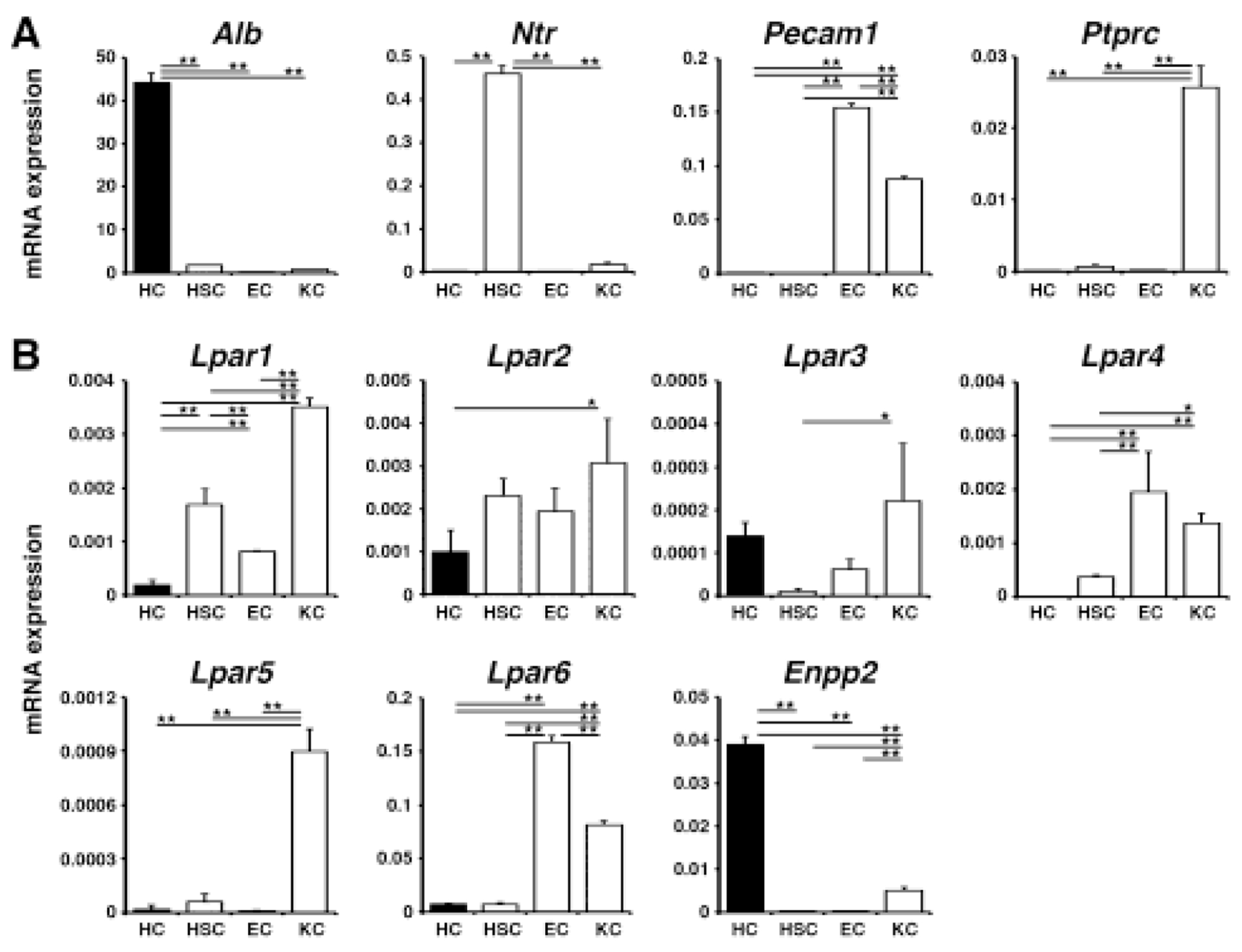
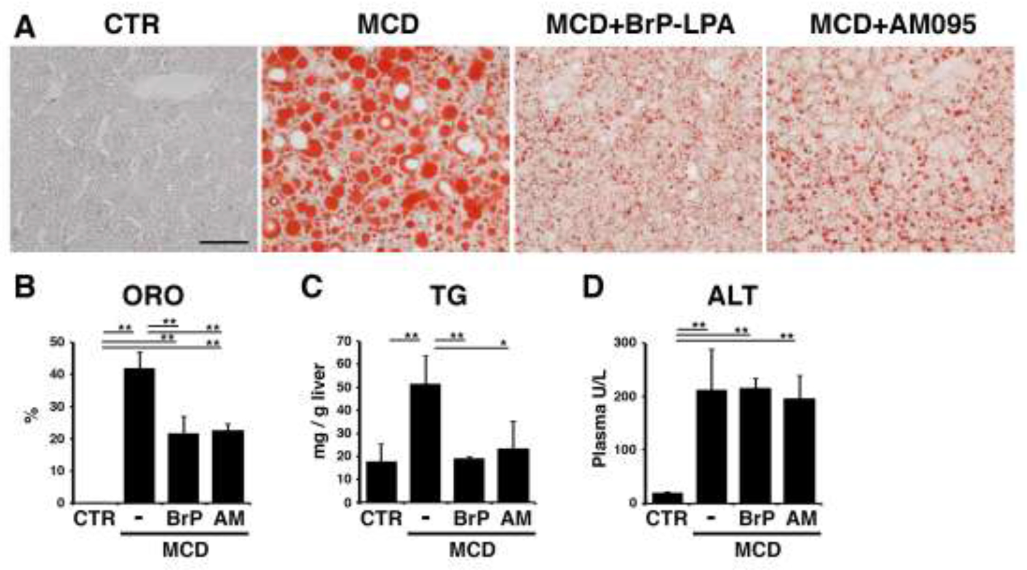
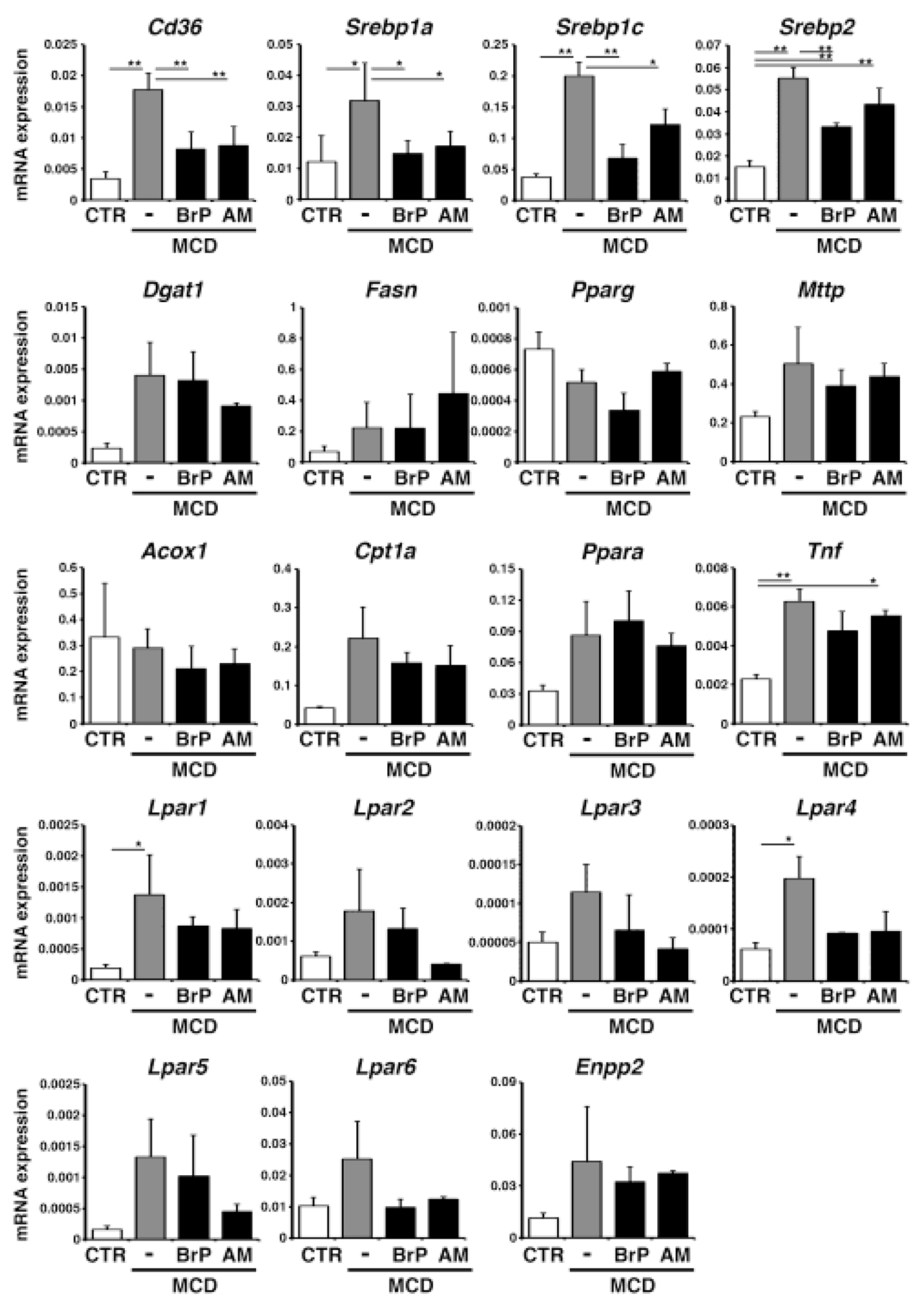
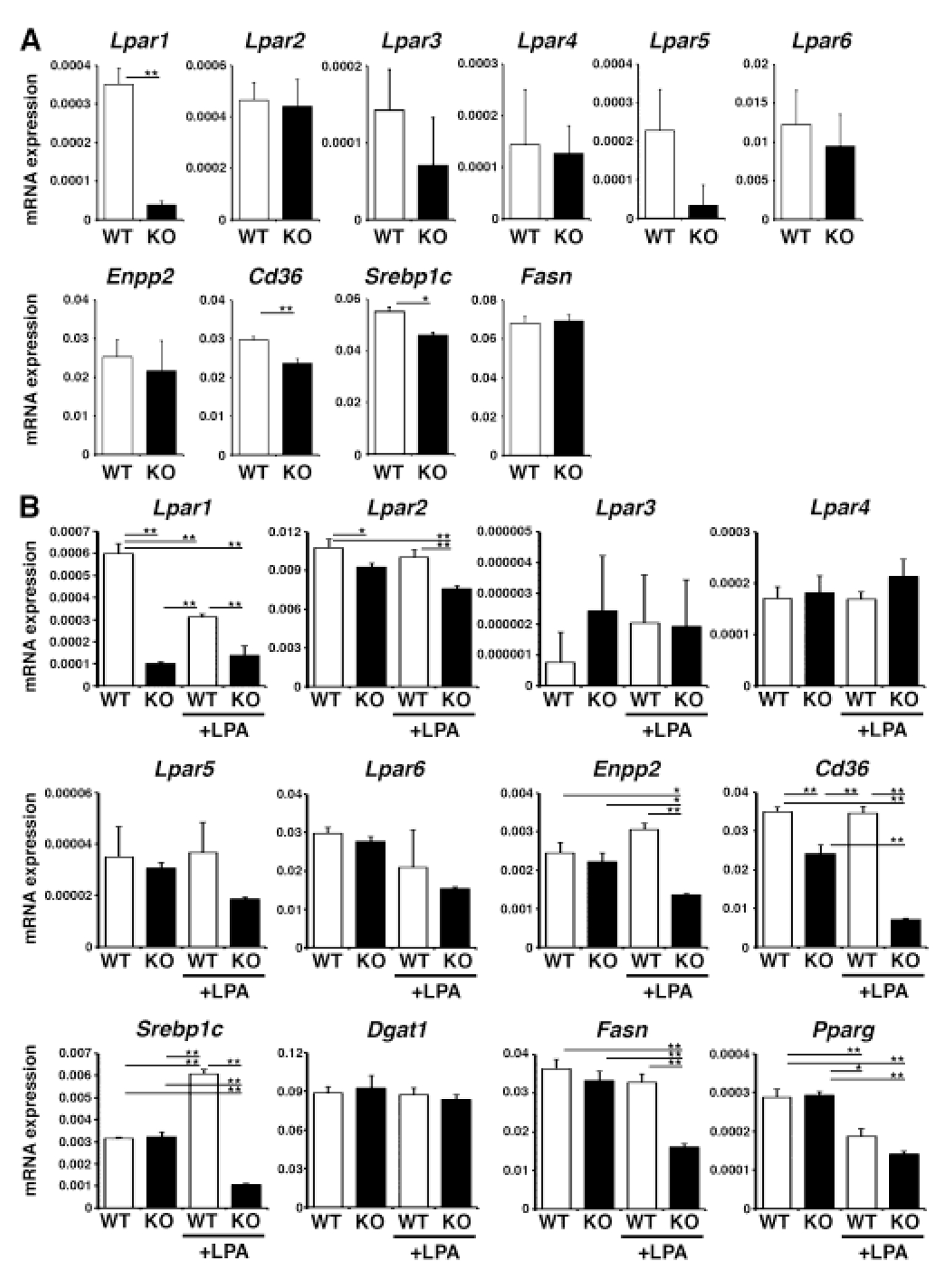
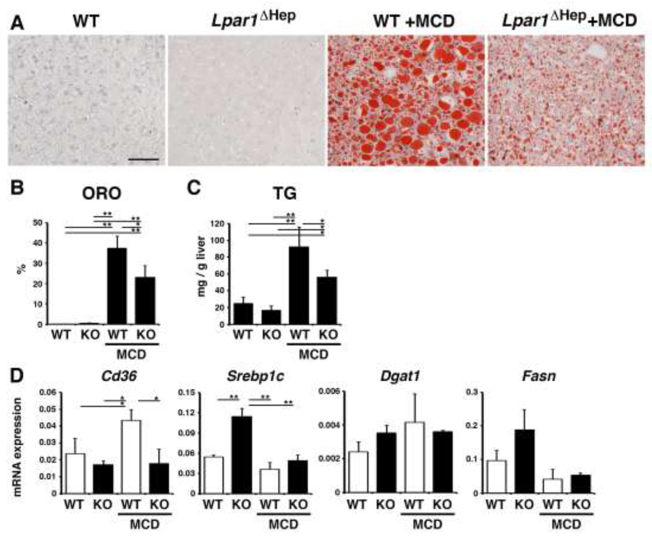
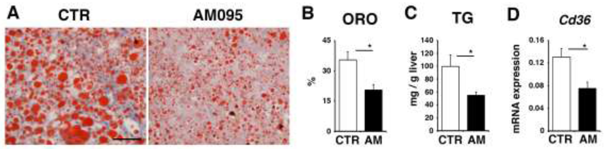
Nonalcoholic steatohepatitis is a major public health concern and is characterized by the accumulation of triglyceride in hepatocytes and inflammation in the liver. Steatosis is caused by dysregulation of the influx and efflux of lipids, lipogenesis, and mitochondrial β-oxidation. Extracellular lysophosphatidic acid (LPA) regulates a broad range of cellular processes in development, tissue injury, and cancer. In the present study, we examined the roles of LPA in steatohepatitis induced by a methionine-choline-deficient (MCD) diet in mice. Hepatocytes express LPA receptor (Lpar) 1-3 mRNAs. Steatosis developed in mice fed the MCD diet was reduced by treatment with inhibitors for pan-LPAR or LPAR1. Hepatocyte-specific deletion of the Lpar1 gene also reduced the steatosis in the MCD model. Deletion of the Lpar1 gene in hepatocytes reduced expression of Cd36, a gene encoding a fatty acid transporter. Although LPA/LPAR1 signaling induces expression of Srebp1 mRNA in hepatocytes, LPA does not fully induce expression of SREBP1-target genes involved in lipogenesis. Human hepatocytes repopulated in chimeric mice are known to develop steatosis and treatment with an LPAR1 inhibitor reduces expression of CD36 mRNA and steatosis. Our data indicate that antagonism of LPAR1 reduces steatosis in mouse and human hepatocytes by down-regulation of Cd36.

Keywords: LPA; LPAR1; Methionine-choline-deficient diet; SREBP1; Steatohepatitis.


Conflict of interest statement

Declaration of competing interest

The authors declare that they have no known competing financial interests or personal relationships that could have appeared to influence the work reported in this paper.

Figures

Fig. 1.
Fig. 1.. Expression profiles of Lpar1-6 and Enpp2 genes in different liver cell types.
QPCR of hepatocytes (HC), hepatic stellate cells (HSC), sinusoidal endothelial cells (EC), and Kupffer cells (KC) isolated from normal mouse livers. (A) Expression of marker genes for each cell type. (B) Expression of Lpar1-6 and Enpp2 mRNAs in different liver cell types. mRNA expression values were normalized against Gapdh. Each value is the mean ± SD of triplicate measurements. *, p < 0.05; **, p <0.01.
Fig. 2.
Fig. 2.. Antagonism of LPAR1 signaling ameliorates MCD-induced steatosis in mice.
Mice fed the MCD diet for 10 days were treated with BrP-LPA (a broad LPAR antagonist), AM095 (an LPAR1 antagonist), or control DMSO on day 3, 6, and 9 (male, n=3 for each group). (A) Oil red O (ORO) staining of the livers. MCD-induced steatosis is ameliorated by BrP-LPA or AM095 treatment. (B) Quantification of ORO staining in each group. (C) Measurement of triglyceride (TG) levels in the livers. (D) Plasma aminotransferase (ALT) levels. *, p < 0.05; **, p < 0.01.
Fig. 3.
Fig. 3.. Antagonism of LPAR1 signaling down-regulates Cd36 mRNA expression in the MCD-fed mouse liver.
Mice fed the MCD diet for 10 days were treated with BrP-LPA (BrP: broad LPAR antagonist), AM095 (AM: LPAR1 antagonist), or control DMSO on day 5, 7, and 9 (male, n=3 for each group). mRNA expression of the liver tissues was analyzed by QPCR. mRNA expression values were normalized against Gapdh. Each value is the mean ± SD of triplicate measurements. *, p < 0.05; **, p<0.01.
Fig. 4.
Fig. 4.. Down-regulation of Cd36 mRNA expression in Lpar1-null hepatocytes.
The Lpar1 gene was deleted in hepatocytes in AlbCre; Lpar1fkox/flox (Lpar1ΔHep) mice. (A) QPCR of hepatocytes isolated from the wild type Lpar1flox/flox (WT) and Lpar1ΔHep (KO) mice. Note the efficient deletion of Lpar1 mRNA in KO. (B) QPCR of cultured hepatocytes treated with LPAfor 2 days. *, p < 0.05; **, p < 0.01.
Fig. 5.
Fig. 5.. Hepatocyte-specific deletion of Lpar1 gene reduces steatosis in mice fed the MCD diet.
Control Lpar1flox/flox (WT) and Lpar1ΔHep (KO) mice were fed with the MCD diet for 10 days (male, n=3 for each group). (A) Oil red O staining of the liver tissues from the WT and Lpar1ΔHep mice fed with the control or MCD diet. After the MCD diet, the Lpar1ΔHep liver shows less accumulation of lipids compared to the WT liver. (B) Quantification of oil red O (ORO) staining in each group. (C) Measurement of triglyceride levels (TG) in the livers. (D) mRNA expression of the liver tissues was analyzed by QPCR. mRNA expression values were normalized against Gapdh. Each value is the mean ± SD of triplicate measurements. *, p < 0.05; **, p < 0.01.
Fig. 6.
Fig. 6.. Reduction of steatosis in chimeric mice bearing human hepatocytes by antagonism of LPAR1
. cDNA-uPA/Scid mice with humanized livers were treated with control DMSO or AM095 every 3 days for a total of 3 times (male, n=3 for each group) and the livers were analyzed. (A) Oil red O staining of the liver tissues from the chimeric mice with AM095 or control DMSO. (B) Quantification of oil red O (ORO) staining. (C) Measurement of triglyceride levels (TG) in the livers. (D) CD36 mRNA expression of the liver tissues analyzed by QPCR. mRNA expression values were normalized against Gapdh. Each value is the mean ± SD of triplicate measurements. *, p < 0.05.

References

    1. Sheka AC, Adeyi O, Thompson J, Hameed B, Crawford PA, Ikramuddin S, Nonalcoholic steatohepatitis: a review, JAMA, 323 (2020) 1175–1183. - PubMed
    1. Schuster S, Cabrera D, Arrese M, Feldstein AE, Triggering and resolution of inflammation in NASH, Nat Rev Gastroenterol Hepatol, 15 (2018) 349–364. - PubMed
    1. Anstee QM, Day CP, The genetics of NAFLD, Nat Rev Gastroenterol Hepatol, 10 (2013) 645–655. - PubMed
    1. Malhi H, Gores GJ, Molecular mechanisms of lipotoxicity in nonalcoholic fatty liver disease, Semin Liver Dis, 28 (2008) 360–369. - PMC - PubMed
    1. Chakravarthy MV, Neuschwander-Tetri BA, The metabolic basis of nonalcoholic steatohepatitis, Endocrinol Diabetes Metab, 3 (2020) e00112. - PMC - PubMed

MeSH terms

LinkOut - more resources