Review
. 2021 Aug 15:427:117527.
doi: 10.1016/j.jns.2021.117527. Epub 2021 Jun 17.

Neurocysticercosis. A frequent cause of seizures, epilepsy, and other neurological morbidity in most of the world

Affiliations
Review

Neurocysticercosis. A frequent cause of seizures, epilepsy, and other neurological morbidity in most of the world

J Bustos et al. J Neurol Sci. .

Abstract

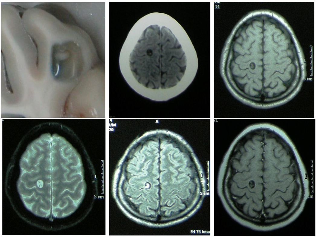
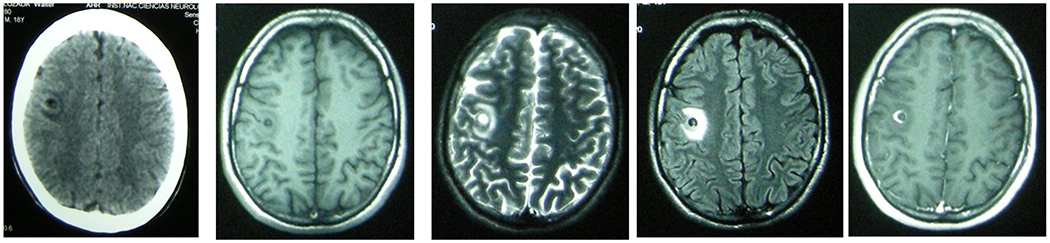
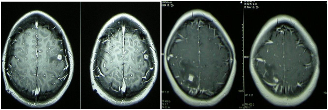
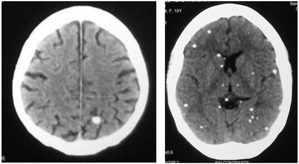
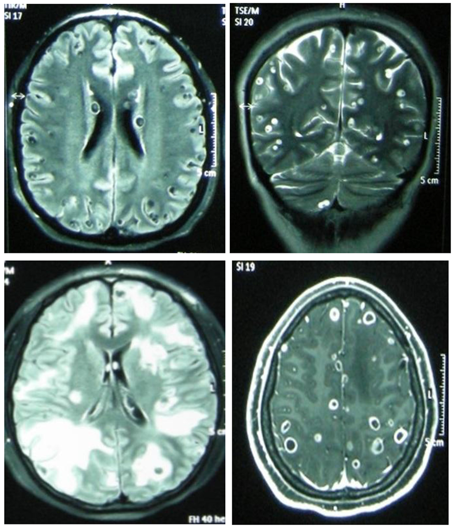
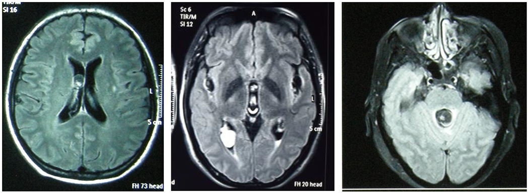
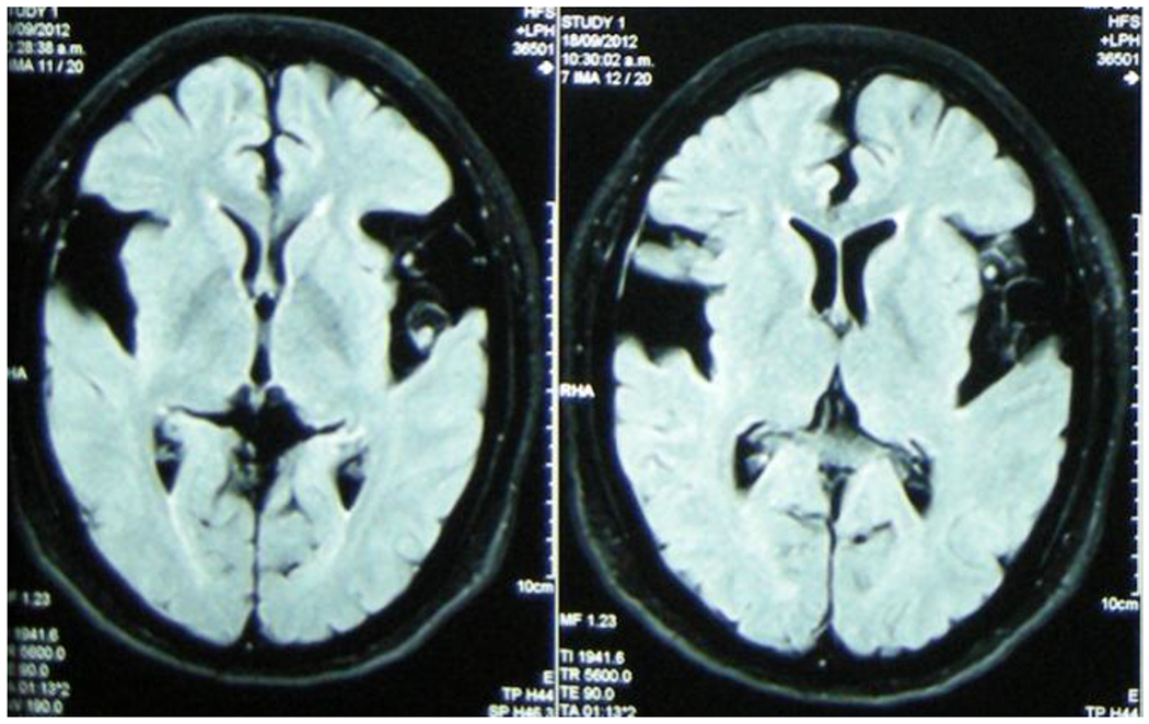
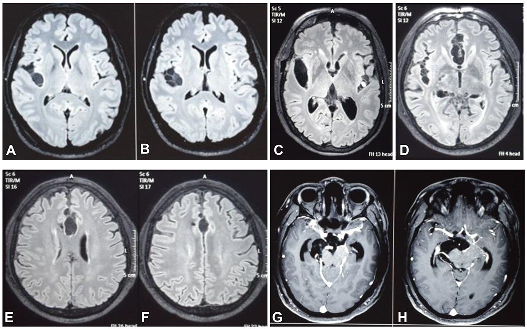
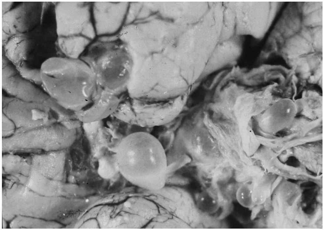
Neurocysticercosis is endemic in most of the world and in endemic areas it accounts for approximately 30% of cases of epilepsy. Appropriate diagnosis and management of neurocysticercosis requires understanding the diverse presentations of the disease since these will vary in regards to clinical manifestation, sensitivity of diagnostic tests, and most importantly, therapeutic approach. This review attempts to familiarize tropical neurology practitioners with the diverse types of neurocysticercosis and the more appropriate management approaches for each.

Keywords: Cysticercosis; Epilepsy; Neurocysticercosis; Taenia solium; Tropical neurology.


Figures

Figure 1,
Figure 1,. Viable cyst without inflammation.
From left to right, upper row: macroscopic view of a cysticercus in the brain of a pig showing the cystic cavity and the scolex; contrast-enhanced CT scan, and non-contrasted T1 MRI. Lower row: non-contrasted T2 and FLAIR, and contrast-enhanced T1 MRI images. Note the absence of contrast enhancement or perilesional edema.
Figure 2,
Figure 2,. Viable cyst with inflammation.
From left to right: contrast-enhanced CT scan, non-contrasted T1, T2 and FLAIR, and contrast-enhanced T1 MRI images. Note the presence of perilesional edema and contrast enhancement.
Figure 3.
Figure 3.
Degenerating cyst (“enhancing lesion”). Contrast-enhanced CT demonstrating a ring enhancing lesion (left) and a nodular enhancing lesion (right), both with perilesional edema. Liquid cyst contents are not noticeable anymore.
Figure 4.
Figure 4.
Single and multiple calcified cysts on non-contrasted CT scan
Figure 5.
Figure 5.
Massive parenchymal NCC, non-encephalitic (above, FLAIR and T2 MRI protocols) and encephalitic (below, FLAIR and post-contrast T1)
Figure 6.
Figure 6.
Ventricular NCC. Left-viable cyst in a lateral ventricle; Center - degenerating cyst in the occipital horn of the right lateral ventricle – note the change in the intensity of signal of the cyst contents; Right - IV ventricle cyst growing towards the aqueduct. All images are non-contrasted MRI, FLAIR protocol.
Figure 7.
Figure 7.
NCC cysts in contact with the subarachnoid space at the Sylvian cisterns, with visible scolices (non-contrasted MRI, FLAIR protocol).
Figure 8.
Figure 8.
Subarachnoid NCC. A - C) Sylvian; D) lower interhemispheric; E,F) upper interhemispheric; G,H) surrounding the brainstem (A to F, non-contrasted MRI FLAIR; G and H, post contrast T1-weighted images).
Figure 9.
Figure 9.
Macroscopic pathology of basal subarachnoid NCC (Reproduced with permission from Garcia HH, Martinez SM (ed). 1996. Taeniasis/cisticercosis por Taenia solium. Editorial Universo, Lima, Peru).
Figure 10.
Figure 10.
Sequential appearance of specific anti-Taenia solium antibodies on enzyme-linked immunoelectrotransfer blot assay using lentil.lectin purified parasite glycoprotein antigens (LLGP-EITB) after experimental pig infection.

References

    1. Garcia HH, Gonzalez AE, Gilman RH. Taenia solium Cysticercosis and Its Impact in Neurological Disease. Clin Microbiol Rev. 2020;33(3). Epub 2020/05/29. - PMC - PubMed
    1. Carod JF, Dorny P. Cysticercosis in Madagascar. J Infect Dev Ctries. 2020;14(9):931–42. Epub 2020/10/09. - PubMed
    1. Mukuku O, Nawej P, Bugeme M, Nduu F, Mawaw PM, Luboya ON. Epidemiology of Epilepsy in Lubumbashi, Democratic Republic of Congo. Neurol Res Int. 2020;2020:5621461. Epub 2020/05/16. - PMC - PubMed
    1. Li T, Chen X, Wang H, Openshaw JJ, Zhong B, Felt SA, et al. High prevalence of taeniasis and Taenia solium cysticercosis in children in western Sichuan, China. Acta Trop. 2019;199:105133. Epub 2019/08/16. - PubMed
    1. Ndimubanzi PC, Carabin H, Budke CM, Nguyen H, Qian YJ, Rainwater E, et al. A systematic review of the frequency of neurocyticercosis with a focus on people with epilepsy. PLoS neglected tropical diseases. 2010;4(11):e870. Epub 2010/11/13. - PMC - PubMed