. 2021 Aug 9;379(2203):20200391.
doi: 10.1098/rsta.2020.0391. Epub 2021 Jun 21.

Pattern formation in drying blood drops

Affiliations

Pattern formation in drying blood drops

Michael J Hertaeg et al. Philos Trans A Math Phys Eng Sci. .

Abstract

Patterns in dried droplets are commonly observed as rings left after spills of dirty water or coffee have evaporated. Patterns are also seen in dried blood droplets and the patterns have been shown to differ from patients afflicted with different medical conditions. This has been proposed as the basis for a new generation of low-cost blood diagnostics. Before these diagnostics can be widely used, the underlying mechanisms leading to pattern formation in these systems must be understood. We analyse the height profile and appearance of dispersions prepared with red blood cells (RBCs) from healthy donors. The red cell concentrations and diluent were varied and compared with simple polystyrene particle systems to identify the dominant mechanistic variables. Typically, a high concentration of non-volatile components suppresses ring formation. However, RBC suspensions display a greater volume of edge deposition when the red cell concentration is higher. This discrepancy is caused by the consolidation front halting during drying for most blood suspensions. This prevents the standard horizontal drying mechanism and leads to two clearly defined regions in final crack patterns and height profile. This article is part of a discussion meeting issue 'A cracking approach to inventing new tough materials: fracture stranger than friction'.

Keywords: blood; coffee ring; diagnostics; droplet drying; drying.


Figures
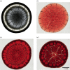
Figure 1.
Figure 1.
Images of droplets dried on untreated glass. Plasma with different concentrations of RBCs: (a) 0 vol.%, (b) 15 vol.%, (c) 30 vol.%, (d) 45 vol.%. Scale bar applies to all images. (Online version in colour.)
Figure 2.
Figure 2.
Schematic showing the drying of a droplet of blood. (a) Early times, similar to process in pure fluids. (b) Mid-late times showing the formation of a consolidated/gelled region with no fluid flow and a reduced evaporation. (Online version in colour.)
Figure 3.
Figure 3.
Profilometry centerline scans of dried 6 μl droplets of RBCs suspended in plasma on untreated glass. Insets show representative images of dried deposits. Effect of RBC concentration in plasma: (a) 0 vol.%, (b) 15 vol.%, (c) 30 vol.% and (d) 45 vol.%. (Online version in colour.)
Figure 4.
Figure 4.
Profilometry centerline scans of dried 6 μl droplets of RBCs suspended in PBS solutions on untreated glass. Insets show representative images of dried deposits. Effect of RBC concentration in PBS: (a) 15 vol.%, (b) 30 vol.% and (c) 45 vol.%. (Online version in colour.)
Figure 5.
Figure 5.
Profilometry centerline scans of dried 6 μl droplets of RBCs suspended in BSA solutions on untreated glass. Insets show representative images of dried deposits. Effect of RBC concentration in BSA: (a) 0 vol.%, (b) 15 vol.%, (c) 30 vol.% and (d) 45 vol.%. (Online version in colour.)
Figure 6.
Figure 6.
Photographs showing the drying of a droplet of whole blood on untreated glass. The approximate diameter of the droplet was 4 mm. (Online version in colour.)
Figure 7.
Figure 7.
Profilometry scans and photographs of dried droplets of two different concentrations of RBCs suspended in PBS solutions on treated glass, controlling contact angle. (a) 15 vol.% and (b) 30 vol.%. (Online version in colour.)

Similar articles

Cited by

References

    1. Peschel O, Kunz SN, Rothschild MA, Mützel E. 2011. Blood stain pattern analysis. Forensic Sci. Med. Pathol. 7, 257–270. (10.1007/s12024-010-9198-1) - DOI - PubMed
    1. Kulyabina TV, Drajevsky RA, Kochubey VI, Zimnyakov DA. 2001. Coherent optical analysis of crystal- like patterns induced by human blood plasma desiccation. Saratov Fall Meeting 2000: Coherent Optics of Ordered and Random Media. Vol. 4242, pp. 282–285.
    1. Chen R, Zhang L, Zang D, Shen W. 2016. Blood drop patterns: formation and applications. Adv. Colloid Interface Sci. 231, 1–14. (10.1016/j.cis.2016.01.008) - DOI - PubMed
    1. Rapis E. 2002. A change in the physical state of a nonequilibrium blood plasma protein film in patients with carcinoma. Tech. Phys. 47, 510–512. (10.1134/1.1470608) - DOI
    1. Brutin D, Sobac B, Loquet B, Sampol J. 2011. Pattern formation in drying drops of blood. J. Fluid Mech. 667, 85–95. (10.1017/S0022112010005070) - DOI

LinkOut - more resources