. 2021 Sep;33(5):884-893.
doi: 10.1177/10406387211025513. Epub 2021 Jun 19.

Oxidative burst and phagocytic activity of phagocytes in canine parvoviral enteritis

Affiliations

Oxidative burst and phagocytic activity of phagocytes in canine parvoviral enteritis

Kelly du Preez et al. J Vet Diagn Invest. 2021 Sep.

Abstract

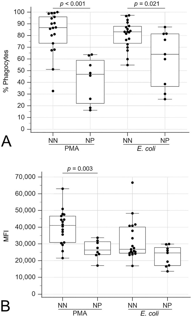
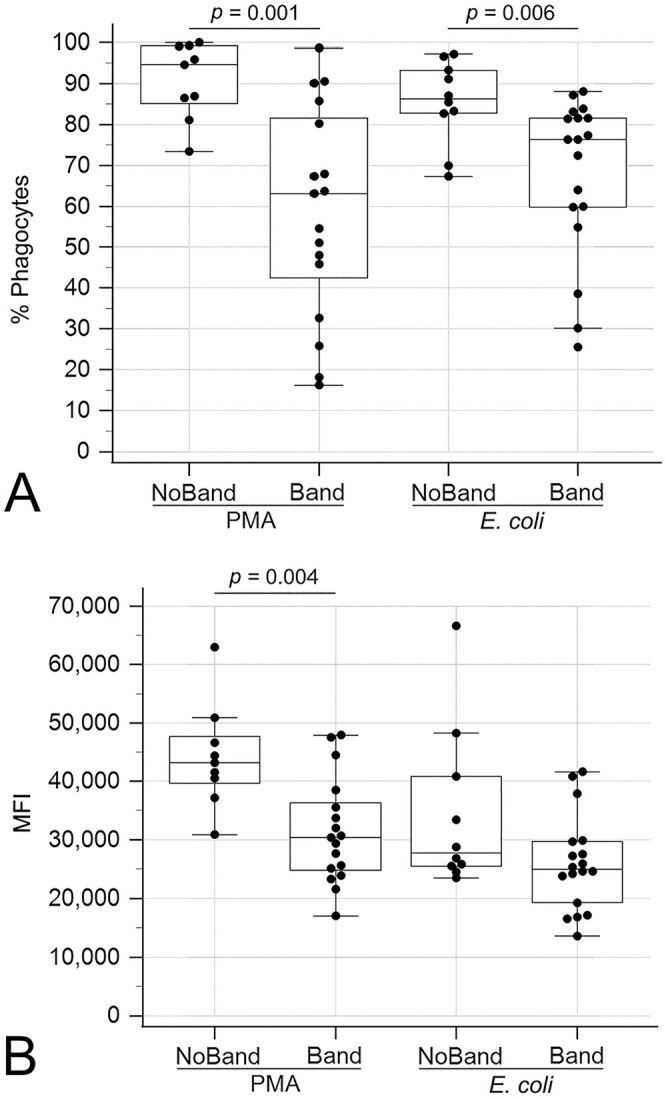
Canine parvoviral enteritis (CPE) is a severe disease characterized by systemic inflammation and immunosuppression. The function of circulating phagocytes (neutrophils and monocytes) in affected dogs has not been fully investigated. We characterized the functional capacity of canine phagocytes in CPE by determining their oxidative burst and phagocytic activities using flow cytometry. Blood was collected from 28 dogs with CPE and 11 healthy, age-matched, control dogs. Oxidative burst activity was assessed by stimulating phagocytes with opsonized Escherichia coli or phorbol 12-myristate 13-acetate (PMA) and measuring the percentage of phagocytes producing reactive oxygen species and the magnitude of this production. Phagocytosis was measured by incubating phagocytes with opsonized E. coli and measuring the percentage of phagocytes containing E. coli and the number of bacteria per cell. Complete blood counts and serum C-reactive protein (CRP) concentrations were also determined. Serum CRP concentration was negatively and positively correlated with segmented and band neutrophil concentrations, respectively. Overall, no differences in phagocyte function were found between dogs with CPE and healthy control dogs. However, infected dogs with neutropenia or circulating band neutrophils had decreased PMA-stimulated oxidative burst activity compared to healthy controls. Additionally, CPE dogs with neutropenia or circulating band neutrophils had decreased PMA- and E. coli-stimulated oxidative burst activity and decreased phagocytosis of E. coli compared to CPE dogs without neutropenia or band neutrophils. We conclude that phagocytes have decreased oxidative burst and phagocytic activity in neutropenic CPE dogs and in CPE dogs with circulating band neutrophils.

Keywords: dogs; flow cytometry; inflammation; phagocyte; phagocytosis; respiratory burst.


Conflict of interest statement

Declaration of conflicting interests: The authors declared no potential conflicts of interest with respect to the research, authorship, and/or publication of this article.

Figures

Figure 1.
Figure 1.
Gating strategy for measuring oxidative burst and phagocytic activities using flow cytometry. A. Live gate (red bracket) on histogram to exclude nonviable cells and bacterial aggregates (blue bracket). B. Forward-scatter (FSC) versus side-scatter (SSC) plot to gate phagocytes (red population). C. Histogram of negative control sample with <3% positive cells (inside blue bracket). D. Example of a histogram from a sample with a population of majority positive cells (inside blue bracket) and some negative cells (outside blue bracket). FITC = fluorescein isothiocyanate.
Figure 2.
Figure 2.
Boxplot comparing the percentage of positive phagocytes undergoing phorbol 12-myristate 13-acetate (PMA)-stimulated oxidative burst between control dogs, neutropenic (NP) dogs with canine parvoviral enteritis (CPE), and CPE dogs with circulating band neutrophils (Band). The box represents the interquartile range, and the bottom and top box margins represent the 25th and 75th interquartiles, respectively; the central line represents the median; the bottom and top whiskers represent the range of the data; and the solid dots represent cases.
Figure 3.
Figure 3.
Boxplots comparing oxidative burst activities between neutropenic (NP) and non-neutropenic (NN) dogs with canine parvoviral enteritis. A. Percentage of positive phagocytes undergoing oxidative burst with phorbol 12-myristate 13-acetate (PMA) or Escherichia coli stimulation. B. Mean fluorescence intensity (MFI) of positive phagocytes undergoing oxidative burst with PMA or E. coli stimulation. The box represents the interquartile range, and the bottom and top box margins represent the 25th and 75th interquartiles, respectively; the central line represents the median; the bottom and top whiskers represent the range of the data; and the symbols represent cases, including outliers that are outside the whiskers.
Figure 4.
Figure 4.
Boxplots comparing phagocytosis between neutropenic (NP) and non-neutropenic (NN) dogs with canine parvoviral enteritis. A. Percentage of cells phagocytizing opsonized Escherichia coli. B. Mean fluorescence intensity (MFI) of cells phagocytizing opsonized E. coli. The box represents the interquartile range, and the bottom and top box margins represent the 25th and 75th interquartiles, respectively; the central line represents the median; the bottom and top whiskers represent the range of the data; and the symbols represent cases, including outliers that are outside the whiskers.
Figure 5.
Figure 5.
Boxplots comparing oxidative burst activities between dogs suffering from canine parvoviral enteritis with circulating band neutrophils (Band) and without circulating band neutrophils (NoBand). A. Percentage of positive phagocytes undergoing oxidative burst with phorbol 12-myristate 13-acetate (PMA) or Escherichia coli stimulation. B. Mean fluorescence intensity (MFI) of positive phagocytes undergoing oxidative burst with PMA or E. coli stimulation. The box represents the interquartile range, and the bottom and top box margins represent the 25th and 75th interquartiles, respectively; the central line represents the median; the bottom and top whiskers represent the range of the data; and the symbols represent cases, including outliers that are outside the whiskers.
Figure 6.
Figure 6.
Boxplot comparing the percentage of cells phagocytizing opsonized Escherichia coli between dogs suffering from canine parvoviral enteritis with circulating band neutrophils (Band) and without circulating band neutrophils (NoBand). The box represents the interquartile range, and the bottom and top box margins represent the 25th and 75th interquartiles, respectively; the central line represents the median; the bottom and top whiskers represent the range of the data; and the symbols represent cases, including outliers that are outside the whiskers.

References

    1. Ahmad M, et al.. In vivo effect of recombinant human granulocyte colony-stimulating factor on phagocytic function and oxidative burst activity in septic neutropenic neonates. Biol Neonate 2004;86:48–54. - PubMed
    1. Allison LN, et al.. Immune function and serum vitamin D in shelter dogs: a case-control study. Vet J 2020;261:1–8. - PubMed
    1. Bardoel BW, et al.. The balancing act of neutrophils. Cell Host Microbe 2014;15:526–536. - PubMed
    1. Bhan C, et al.. Role of cellular events in the pathophysiology of sepsis. Inflamm Res 2016;65:853–868. - PubMed
    1. Biswas SK, et al.. Endotoxin tolerance: new mechanisms, molecules and clinical significance. Trends Immunol 2009;30:475–487. - PubMed