. 2021 Jun 21;8(1):37.
doi: 10.1186/s40779-021-00333-4.

RIG-I, a novel DAMPs sensor for myoglobin activates NF-κB/caspase-3 signaling in CS-AKI model

Affiliations

RIG-I, a novel DAMPs sensor for myoglobin activates NF-κB/caspase-3 signaling in CS-AKI model

Peng-Tao Wang et al. Mil Med Res. .

Abstract

Background: Acute kidney injury (AKI) is the main life-threatening complication of crush syndrome (CS), and myoglobin is accepted as the main pathogenic factor. The pattern recognition receptor retinoicacid-inducible gene I (RIG-I) has been reported to exert anti-viral effects function in the innate immune response. However, it is not clear whether RIG-I plays a role in CS-AKI. The present research was carried out to explore the role of RIG-I in CS-AKI.

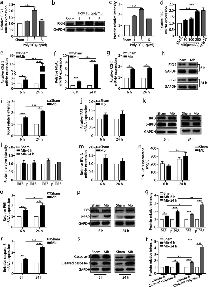
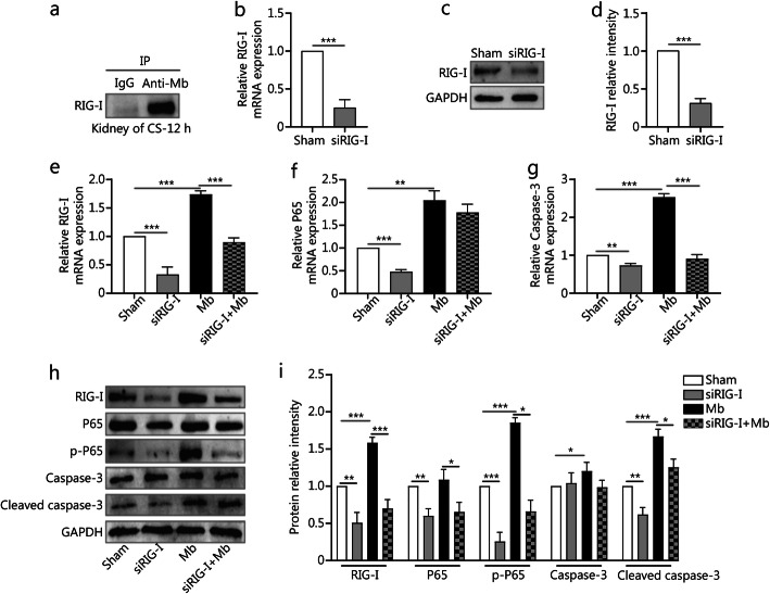
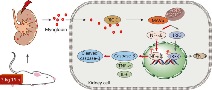
Methods: Sprague-Dawley rats were randomly divided into two groups: the sham and CS groups (n = 12). After administration of anesthesia, the double hind limbs of rats in the CS group were put under a pressure of 3 kg for 16 h to mimic crush conditions. The rats in both groups were denied access to food and water. Rats were sacrificed at 12 h or 36 h after pressure was relieved. The successful establishment of the CS-AKI model was confirmed by serum biochemical analysis and renal histological examination. In addition, RNA sequencing was performed on rat kidney tissue to identify molecular pathways involved in CS-AKI. Furthermore, NRK-52E cells were treated with 200 μmol/L ferrous myoglobin to mimic CS-AKI at the cellular level. The cells and cell supernatant samples were collected at 6 h or 24 h. Small interfering RNAs (siRNA) was used to knock down RIG-I expression. The relative expression levels of molecules involved in the RIG-I pathway in rat kidney or cells samples were measured by quantitative Real-time PCR (qPCR), Western blotting analysis, and immunohistochemistry (IHC) staining. Tumor necrosis factor-α (TNF-α) was detected by ELISA. Co-Immunoprecipitation (Co-IP) assays were used to detect the interaction between RIG-I and myoglobin.

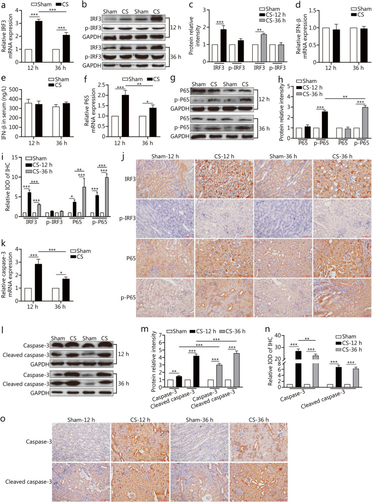
Results: RNA sequencing of CS-AKI rat kidney tissue revealed that the different expression of RIG-I signaling pathway. qPCR, Western blotting, and IHC assays showed that RIG-I, nuclear factor kappa-B (NF-κB) P65, p-P65, and the apoptotic marker caspase-3 and cleaved caspase-3 were up-regulated in the CS group (P < 0.05). However, the levels of interferon regulatory factor 3 (IRF3), p-IRF3 and the antiviral factor interferon-beta (IFN-β) showed no significant changes between the sham and CS groups. Co-IP assays showed the interaction between RIG-I and myoglobin in the kidneys of the CS group. Depletion of RIG-I could alleviate the myoglobin induced expression of apoptosis-associated molecules via the NF-κB/caspase-3 axis.

Conclusion: RIG-I is a novel damage-associated molecular patterns (DAMPs) sensor for myoglobin and participates in the NF-κB/caspase-3 signaling pathway in CS-AKI. In the development of CS-AKI, specific intervention in the RIG-I pathway might be a potential therapeutic strategy for CS-AKI.

Keywords: Acute kidney injury; Crush syndrome; Damage-associated molecular patterns; Myoglobin; Nuclear factor kappa-B/caspase-3; Retinoic acid-inducible gene I.


Conflict of interest statement

The authors declare that there are no competing interests.

Figures

Fig. 1
Fig. 1
The expression of RIG-I in the CS-AKI rat model. DEGs: Differentially expressed genes. a–d. Serum levels of CK, Scr, BUN, and Mb. e–f. qPCR analysis of renal KIM-1 and NGAL expression. g. HE and PAS staining of kidney tissues (scale bar: 100 μm). h. Renal tubular injury score, as determined by calculating the percentage of tubules that displayed tubular dilation, cast formation and tubular necrosis: 0, normal; 1, ≤10%; 2, 10–25%; 3, 26–50%; 4, 51–75%; 5, ≥75%. i. DEGs between the sham and CS groups, as identified by RNA sequencing. j. The 10 signaling pathways, as identified by KEGG pathway analysis. k–o. qPCR, Western blotting and IHC analyses (scale bar: 100 μm) of renal RIG-I expression. Data are expressed as the mean ± SD (n = 6). *P < 0.05, **P < 0.01, ***P < 0.001, two-way ANOVA followed by the Bonferroni’s multiple comparisons test
Fig. 2
Fig. 2
The expressions of downstream molecules involved in RIG-I signaling in the CS-AKI rat model. IRF3: Interferon regulatory factor 3. a–c. qPCR, and Western blotting analyses expression levels of renal IRF3. Fig. c is the quantification of Fig. b. d. qPCR analyses of renal IFN-β expression. e. Serum levels of IFN-β by ELISA. f–h. qPCR, and Western blotting analyses renal P65 and p-P65 expression. Fig. h is the quantification of Fig. g. i–j. IHC analyses renal IRF3, p-IRF3, P65, and p-P65 expression after relieving the pressure at 12 h and 36 h (scale bar: 100 μm). Fig. i is the quantification of IHC staining. k–m. qPCR, and Western blotting analyses renal caspase-3 and cleaved caspase-3 expression. Fig. m is the quantification of Fig. l. n–o. IHC analyses renal caspase-3 and cleaved caspase-3 expression (scale bar: 100 μm), Fig. n is the quantification of Fig. o; Data are expressed as the mean ± SD. *P < 0.05, **P < 0.01, ***P < 0.001, two-way ANOVA followed by the Bonferroni’s multiple comparisons test
Fig. 3
Fig. 3
The molecules involved in RIG-I signaling in the CS-AKI cell model. a–c. qPCR, and Western blotting analyses the RIG-I expression after NRK-52E cells treatment with 1, 3 or 6 μg/ml Poly I:C for 24 h respectively. Fig. c is the quantification of Fig. b. d. qPCR analyses RIG-I expression activated by different concentrations of ferrous myoglobin. e–f. qPCR analyses cells KIM-1 and NGAL expression after treatment with 200 μmol/L ferrous myoglobin at 6 h and 24 h separately. g-i. qPCR, and Western blotting analyses RIG-I expression. Fig. i is the quantification of Fig. h. j–l. qPCR, and Western blotting analyses IRF3 and p-IRF3 expression. Fig. l is the quantification of Fig. k. m. qPCR analyses IFN-β expression. n. Cell supernatant levels of IFN-β by ELISA. o–q. qPCR, and Western blotting analyses P65 and p-P65 expression. Fig. q is the quantification of Fig. p. r–t. qPCR, and Western blotting analyses caspase-3 and cleaved caspase-3 expression. Fig. t is the quantification of Fig. s. Data are expressed as the mean ± SD. *P < 0.05, **P < 0.01, ***P < 0.001, two-way ANOVA followed by the Bonferroni’s multiple comparisons test or one-way ANOVA followed by the Brown–Forsythe multiple comparisons test, respectively
Fig. 4
Fig. 4
Knocking down RIG-I gene could alleviate the myoglobin induced NF-κB/caspase-3 axis activation. a. Co-IP showed the interaction between RIG-I and myoglobin in the rat kidney of the CS group. b–d. qPCR, and Western blotting analyses RIG-I expression after NRK-52E cells treatment with siRIG-I. Fig. d is the quantification of Fig. c. e–i. qPCR, and Western blotting analyses cells RIG-I, IRF3, p-IRF3, P65, p-P65, caspase-3, and cleaved caspase-3 expression after treatment with ferrous myoglobin and siRIG-I. Fig. i is the quantification of Fig. h. Data are expressed as the mean ± SD. *P < 0.05, **P < 0.01, ***P < 0.001, unpaired t-test or by one-way ANOVA followed by the Brown–Forsythe multiple comparisons test, respectively
Fig. 5
Fig. 5
Schematic drawing of the novel RIG-I signaling pathway in CS-AKI. In the crush syndrome related acute kidney injury (CS-AKI) rat model, muscle tissue releases large amounts of myoglobin to injure the kidney. Myoglobin binds to RIG-I after endocytosis, then mainly activate NK-κB/caspase-3 axis, not the classical IRF-3/IFN-β signaling pathway in CS-AKI rat kidney. In this process, the expression of pro-inflammation cytokines IL-6 and TNF-α are also increased to some degree

References

    1. Zhou XL, Ni SZ, Xiong D, Cheng XQ, Xu P, Zhao Y. Fluid resuscitation with preventive peritoneal dialysis attenuates crush injury-related acute kidney injury and improves survival outcome. Scand J Trauma Resusc Emerg Med. 2019;27(1):68. doi: 10.1186/s13049-019-0644-0. - DOI - PMC - PubMed
    1. Kadıoğlu E, Tekşen Y, Koçak C, Koçak FE. Beneficial effects of bardoxolone methyl, an Nrf2 activator, on crush-related acute kidney injury in rats. Eur J Trauma Emerg Surg. 2021;47(1):241–250. doi: 10.1007/s00068-019-01216-z. - DOI - PubMed
    1. Scapellato S, Maria S, Castorina G, Sciuto G. Crush syndrome. Minerva Chir. 2007;62(4):285–292. - PubMed
    1. Li N, Wang X, Wang P, Fan H, Hou S, Gong Y. Emerging medical therapies in crush syndrome - progress report from basic sciences and potential future avenues. Ren Fail. 2020;42(1):656–666. doi: 10.1080/0886022X.2020.1792928. - DOI - PMC - PubMed
    1. Omrani H, Najafi I, Bahrami K, Najafi F, Safari S. Acute kidney injury following traumatic rhabdomyolysis in Kermanshah earthquake victims; a cross-sectional study. Am J Emerg Med. 2021;40:127–132. doi: 10.1016/j.ajem.2020.01.043. - DOI - PubMed

Publication types