Review
. 2021 Oct;46(10):832-847.
doi: 10.1016/j.tibs.2021.05.007. Epub 2021 Jun 18.

Lamin post-translational modifications: emerging toggles of nuclear organization and function

Affiliations
Review

Lamin post-translational modifications: emerging toggles of nuclear organization and function

Laura A Murray-Nerger et al. Trends Biochem Sci. 2021 Oct.

Abstract

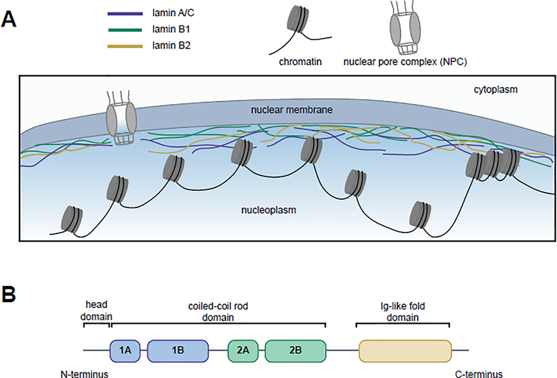
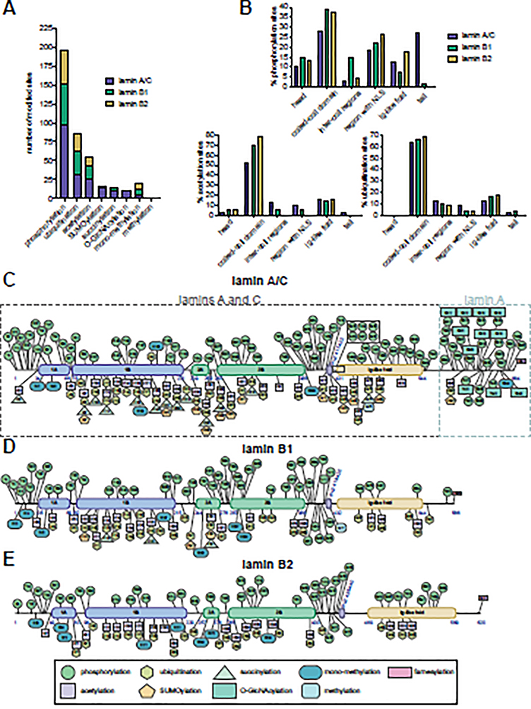
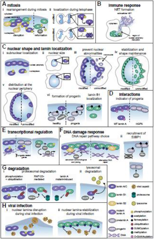
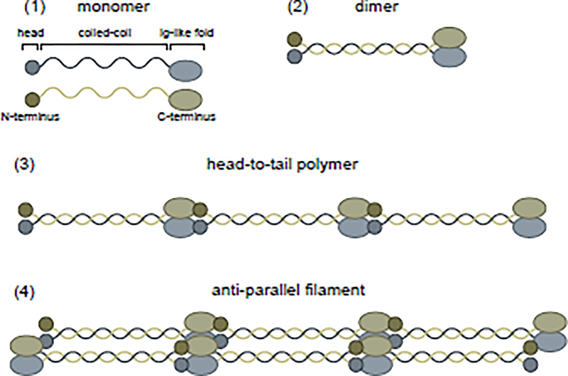
Nuclear lamins are ancient type V intermediate filaments with diverse functions that include maintaining nuclear shape, mechanosignaling, tethering and stabilizing chromatin, regulating gene expression, and contributing to cell cycle progression. Despite these numerous roles, an outstanding question has been how lamins are regulated. Accumulating work indicates that a range of lamin post-translational modifications (PTMs) control their functions both in homeostatic cells and in disease states such as progeria, muscular dystrophy, and viral infection. Here, we review the current knowledge of the diverse types of PTMs that regulate lamins in a site-specific manner. We highlight methods that can be used to characterize lamin PTMs whose functions are currently unknown and provide a perspective on the future of the lamin PTM field.

Keywords: PTMs; acetylation; farnesylation; lamins; phosphorylation; ubiquitination.


Conflict of interest statement

Declaration of interests The authors declare no competing interests.

Figures

Figure 1.
Figure 1.. Lamins’ localization to the nuclear periphery and functional domains.
(A) The human lamins, lamin A/C (blue), lamin B1 (green), and lamin B2 (yellow), assemble into a network at the inner nuclear periphery where they serve to maintain nuclear shape and interact with both euchromatic and heterochromatic regions of DNA. (B) Lamins have three domains: a head domain, a coiled-coil rod domain (composed of four sub-domains) that mediates interactions with other lamina proteins, and an Ig-like fold domain that mediates interactions with non-lamina proteins.
Figure 2.
Figure 2.. The expanding landscape of human lamin post-translational modifications.
(A) Number of modifications on the human lamins per PTM type. (B) Percentage of phosphorylation (upper), acetylation (lower left), and ubiquitination (lower right) sites in the different lamin domains. (C-E) Human lamin PTMs documented on PhosphoSitePlus and/or discussed in this review are indicated for (C) lamin A/C (amino acids shared between lamins A and C and those unique to lamin A are indicated by dashed boxes), (D) lamin B1, and (E) lamin B2. The first and last amino acids of the different domains and the amino acids for the nuclear localization signal (NLS) are indicated in blue.
Figure 3.
Figure 3.. Post-translational modifications regulate lamin functions.
Mature lamin A/C (A/C), lamin A (A), lamin B1 (B1), lamin B2 (B2). (A) (i) During mitosis, lamin phosphorylation promotes nuclear periphery disruption. PP1-mediated B1 dephosphorylation facilitates lamina reformation after mitosis. (ii) At telophase, SUMOylation promotes A dephosphorylation and nuclear relocalization. (B) B1 phosphorylation disrupts the nuclear periphery, releasing chromatin for NET formation by neutrophils. (C) (i) A/C phosphorylation determines sub-nuclear localization. (ii) A phosphorylation maintains nuclear size. (iii) A/C acetylation prevents nuclear deformations. (iv) B1 methylation maintains its localization and nuclear shape. (v) A SUMOylation maintains lamin spacing. (vi) A maturation requires Zmpste24-mediated cleavage of prelamin A and farnesylation removal, which are absent in progeria. Farnesylation promotes proper B1 localization. (D) O-GlcNAcylation decorates wild type A but not progerin. (E) A/C phosphorylation enables its association with enhancers, which is altered by progerin. B1 phosphorylation releases Oct-1. (F) (i) In homeostasis, B1 acetylation increases and decreases associations with lamins and chromatin, respectively. Upon DNA damage, this reduced chromatin association modulates selection of DNA repair pathways. (ii) Farnesylation status of prelamin A impacts 53BP1 recruitment to damaged DNA. (G) (i) A phosphorylation promotes its ubiquitin-mediated degradation, and RNF123-mediated A and B1 ubiquitination induces their degradation. A mutants present in Emery-Dreifuss muscular dystrophy promote B1 ubiquitination-mediated degradation. (ii) Smurf2-mediated ubiquitination is implicated in lysosome-mediated A degradation. (H) (i) A/C phosphorylation during viral infection induces lamina disruption, facilitating viral capsid nuclear egress. (ii) During herpesvirus infection, B1 acetylation inhibits lamina disruption and viral capsid egress.
Figure I.
Figure I.. Lamins assemble into filaments.
Lamins, initially synthesized as monomers (1), assemble into intermediate filaments by first dimerizing (2) and then forming head-to-tail polymers (3). These polymers assemble in an anti-parallel manner into filaments (4).
Figure II.
Figure II.. Methods to detect and functionally characterize lamin PTMs.
(A) Lamin PTMs can be identified by MS using several experimental workflows, including whole proteome analyses (green), PTM-specific enrichment by biochemical or immunoaffinity purification methods (blue), or lamin-specific enrichment by immunoaffinity purification (IP) of a particular lamin (purple). (B) Considerations when characterizing lamin PTMs are (1) validation and quantification, (2) determination of biological function, and (3) assessment of enzymatic regulation. Targeted MS, including selected reaction monitoring (SRM) and parallel reaction monitoring (PRM), provides the means for confirming and accurately quantifying the levels of a site-specific PTM. Biological function can be determined by employing site-directed mutagenesis of the lamin of interest combined with functional assays that test different aspects of lamin biology (examples of functions and experimental techniques are listed). As many enzymes may regulate the same PTM, employing motif and subcellular localization computational analyses (examples of databases/tools are listed) can narrow the focus on which enzyme(s) may work on a particular PTM. The ability of the enzyme(s) to modify that site can then be tested using in vitro (tan) and cell culture and in vivo experiments (blue). Examples of the consequences on PTM levels of an enzyme that adds (teal) or removes (orange) a given PTM are shown.

References

    1. Lin F and Worman HJ (1995) Structural organization of the human gene (LMNB1) encoding nuclear lamin B1. Genomics 27, 230–236 - PubMed
    1. Lin F and Worman HJ (1993) Structural organization of the human gene encoding nuclear lamin A and nuclear lamin C. J. Biol. Chem. 268, 16321–16326 - PubMed
    1. Fisher DZ et al. (1986) cDNA sequencing of nuclear lamins A and C reveals primary and secondary structural homology to intermediate filament proteins. Proc. Natl. Acad. Sci. U. S. A. 83, 6450–6454 - PMC - PubMed
    1. McKeon FD et al. (1986) Homologies in both primary and secondary structure between nuclear envelope and intermediate filament proteins. Nature 319, 463–468 - PubMed
    1. Broers JLV et al. (2006) Nuclear lamins: Laminopathies and their role in premature ageing. Physiol. Rev. 86, 967–1008 - PubMed

Publication types

LinkOut - more resources