

ECG Paper Record Digitization and Diagnosis Using Deep Learning
- PMID: 34149335
- PMCID: PMC8204064
- DOI: 10.1007/s40846-021-00632-0
ECG Paper Record Digitization and Diagnosis Using Deep Learning
Abstract
Purpose: Electrocardiogram (ECG) is one of the most essential tools for detecting heart problems. Till today most of the ECG records are available in paper form. It can be challenging and time-consuming to manually assess the ECG paper records. Hence, automated diagnosis and analysis are possible if we digitize such paper ECG records.
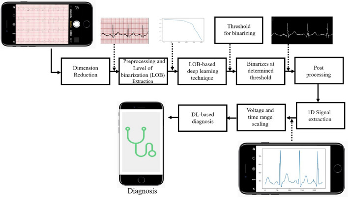
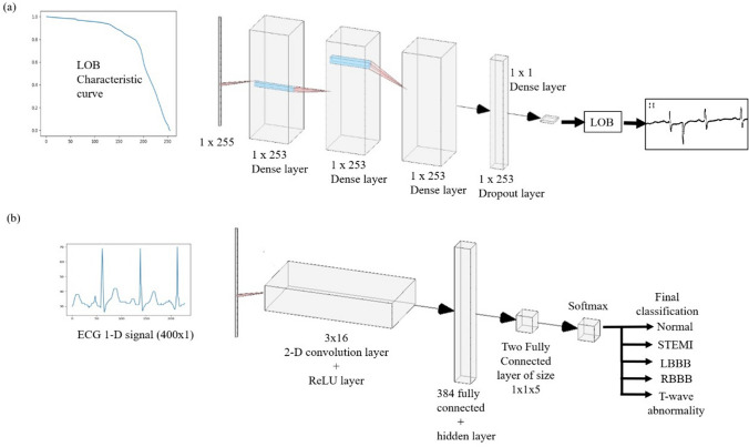
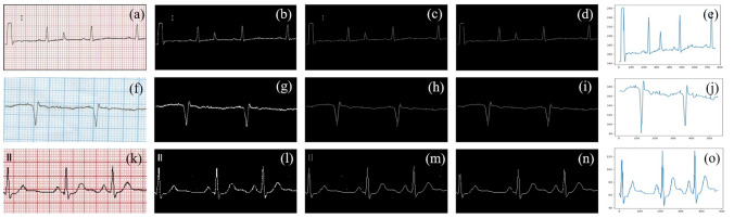
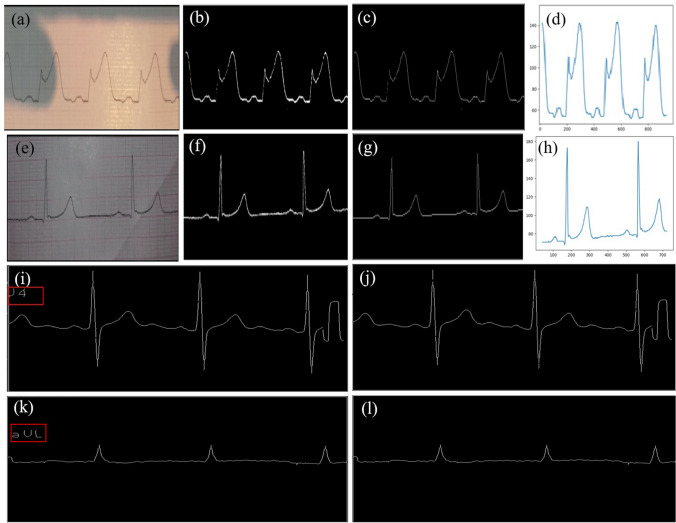
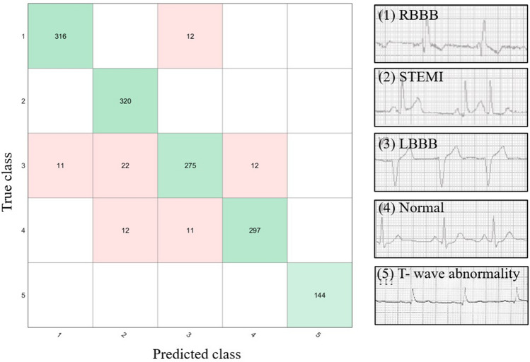
Methods: The proposed work aims to convert ECG paper records into a 1-D signal and generate an accurate diagnosis of heart-related problems using deep learning. Camera-captured ECG images or scanned ECG paper records are used for the proposed work. Effective pre-processing techniques are used for the removal of shadow from the images. A deep learning model is used to get a threshold value that separates ECG signal from its background and after applying various image processing techniques threshold ECG image gets converted into digital ECG. These digitized 1-D ECG signals are then passed to another deep learning model for the automated diagnosis of heart diseases into different classes such as ST-segment elevation myocardial infarction (STEMI), Left Bundle Branch Block (LBBB), Right Bundle Branch Block (RBBB), and T-wave abnormality.
Results: The accuracy of deep learning-based binarization is 97%. Further deep learning-based diagnosis approach of such digitized paper ECG records was having an accuracy of 94.4%.
Conclusions: The digitized ECG signals can be useful to various research organizations because the trends in heart problems can be determined and diagnosed from preserved paper ECG records. This approach can be easily implemented in areas where such expertise is not available.
Supplementary information: The online version contains supplementary material available at 10.1007/s40846-021-00632-0.
Keywords: Deep learning; Diagnosis; Digitization; Paper ECG.
© Taiwanese Society of Biomedical Engineering 2021.
Conflict of interest statement
Conflict of interestThe authors declare that they have no conflict of interest.
Figures
References
-
- Biswas, B., Bhattacharya, U., & Chaudhuri, B. B. (2014). In: Proceedings of the 22nd International Conference on Pattern Recognition, pp. 3008–3013.
-
- Su B, Lu S, Tan CL. IEEE Transactions on Image Processing. 2012;22(4):1408. - PubMed
-
- Sauvola J, Pietikäinen M. Pattern Recognition. 2000;33(2):225. doi: 10.1016/S0031-3203(99)00055-2. - DOI
-
- Swamy, P., Jayaraman, S., & Chandra, M. G. (2010). In: Proceedings of the International Conference on Bioinformatics and Biomedical Technology, pp. 400–403.
-
- Mallawaarachchi, S., Perera, M. P. N., & Nanayakkara, N. D. (2014). In: Proceedings of the IEEE Conference on Biomedical Engineering and Sciences (IECBES), pp. 868–873.
LinkOut - more resources
Full Text Sources
Other Literature Sources