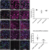
Discovery of a Highly Conserved Peptide in the Iron Transporter Melanotransferrin that Traverses an Intact Blood Brain Barrier and Localizes in Neural Cells
- PMID: 34149342
- PMCID: PMC8212695
- DOI: 10.3389/fnins.2021.596976
Discovery of a Highly Conserved Peptide in the Iron Transporter Melanotransferrin that Traverses an Intact Blood Brain Barrier and Localizes in Neural Cells
Abstract
The blood-brain barrier (BBB) hinders the distribution of therapeutics intended for treatment of diseases of the brain. Our previous studies demonstrated that that a soluble form of melanotransferrin (MTf; Uniprot P08582; also known as p97, MFI2, and CD228), a mammalian iron-transport protein, is an effective carrier for delivery of drug conjugates across the BBB into the brain and was the first BBB targeting delivery system to demonstrate therapeutic efficacy within the brain. Here, we performed a screen to identify peptides from MTf capable of traversing the BBB. We identified a highly conserved 12-amino acid peptide, termed MTfp, that retains the ability to cross the intact BBB intact, distributes throughout the parenchyma, and enter endosomes and lysosomes within neurons, astrocytes and microglia in the brain. This peptide may provide a platform for the transport of therapeutics to the CNS, and thereby offers new avenues for potential treatments of neuropathologies that are currently refractory to existing therapies.
Keywords: MTfp; blood-brain barrer; drug delivery and targeting; glial targeting; melanotransferrin; microglial targeting; neuronal targeting; peptide transport.
Copyright © 2021 Singh, Eyford, Abraham, Munro, Choi, Okon, Vitalis, Gabathuler, Lu, Pfeifer, Tian and Jefferies.
Conflict of interest statement
Bioasis Technologies Inc., (BTI) is a University of British Columbia start-up company. TZV, RG, and MMT were employees and equity holders in BTI at the time this work was undertaken. WAJ was the founding scientist and an equity holder in BTI at the time this work was undertaken. The funding sources had no influence in the study design, data collection, analysis or interpretation of data, in the writing of the manuscript or on the decision to submit for publication. The remaining authors declare that the research was conducted in the absence of any commercial or financial relationships that could be construed as a potential conflict of interest.
Figures


Similar articles
-
A Nanomule Peptide Carrier Delivers siRNA Across the Intact Blood-Brain Barrier to Attenuate Ischemic Stroke.Front Mol Biosci. 2021 Mar 26;8:611367. doi: 10.3389/fmolb.2021.611367. eCollection 2021. Front Mol Biosci. 2021. PMID: 33869275 Free PMC article.
-
Unlocking the potential of melanotransferrin (CD228): implications for targeted drug development and novel therapeutic avenues.Expert Opin Ther Targets. 2024 Dec;28(12):1117-1129. doi: 10.1080/14728222.2024.2441705. Epub 2024 Dec 19. Expert Opin Ther Targets. 2024. PMID: 39676256 Review.
-
A unique carrier for delivery of therapeutic compounds beyond the blood-brain barrier.PLoS One. 2008 Jun 25;3(6):e2469. doi: 10.1371/journal.pone.0002469. PLoS One. 2008. PMID: 18575595 Free PMC article.
-
Directing adenovirus across the blood-brain barrier via melanotransferrin (P97) transcytosis pathway in an in vitro model.Gene Ther. 2007 Mar;14(6):523-32. doi: 10.1038/sj.gt.3302888. Epub 2006 Nov 30. Gene Ther. 2007. PMID: 17167498
-
Blood-brain barrier transport machineries and targeted therapy of brain diseases.Bioimpacts. 2016;6(4):225-248. doi: 10.15171/bi.2016.30. Epub 2016 Dec 5. Bioimpacts. 2016. PMID: 28265539 Free PMC article. Review.
Cited by
-
Advances in research on biomaterials and stem cell/exosome-based strategies in the treatment of traumatic brain injury.Acta Pharm Sin B. 2025 Jul;15(7):3511-3544. doi: 10.1016/j.apsb.2025.05.010. Epub 2025 May 21. Acta Pharm Sin B. 2025. PMID: 40698148 Free PMC article. Review.
-
MFI2 upregulation promotes malignant progression through EGF/FAK signaling in oral cavity squamous cell carcinoma.Cancer Cell Int. 2023 Jun 12;23(1):112. doi: 10.1186/s12935-023-02956-0. Cancer Cell Int. 2023. PMID: 37309001 Free PMC article.
-
Liver-directed gene therapy corrects neurologic disease in a murine model of mucopolysaccharidosis type I-Hurler.Mol Ther Methods Clin Dev. 2022 Apr 19;25:370-381. doi: 10.1016/j.omtm.2022.04.010. eCollection 2022 Jun 9. Mol Ther Methods Clin Dev. 2022. PMID: 35573046 Free PMC article.
-
Changes in Whey Proteome between Mediterranean and Murrah Buffalo Colostrum and Mature Milk Reflect Their Pharmaceutical and Medicinal Value.Molecules. 2022 Feb 27;27(5):1575. doi: 10.3390/molecules27051575. Molecules. 2022. PMID: 35268677 Free PMC article.
-
Melanotransferrin (MELTF, MFI2, CD228) Expression Attenuates Malignant Melanoma Progression in the A375-Luc2 Murine Metastasis Model and Human Patients.J Invest Dermatol. 2024 Dec;144(12):2820-2823.e6. doi: 10.1016/j.jid.2024.05.028. Epub 2024 Jun 21. J Invest Dermatol. 2024. PMID: 38909843 No abstract available.
References
-
- Alemany R., Vila M. R., Franci C., Egea G., Real F. X., Thomson T. M. (1993). Glycosyl phosphatidylinositol membrane anchoring of melanotransferrin (p97): apical compartmentalization in intestinal epithelial cells. J. Cell Sci. 104 1155–1162. - PubMed
-
- Baker E. N., Baker H. M., Smith C. A., Stebbins M. R., Kahn M., Hellstrom K. E., et al. (1992). Human melanotransferrin (p97) has only one functional iron-binding site. FEBS Lett. 298 215–218. - PubMed
-
- Baker_Lab Robetta is a Protein Structure Prediction Service that is Continually Evaluated Through CAMEO. Seattle, WA: Baker_Lab. (accessed 2015).
Grants and funding
LinkOut - more resources
Full Text Sources
Other Literature Sources
Miscellaneous

