Review
. 2021 Jun 2:12:689083.
doi: 10.3389/fphys.2021.689083. eCollection 2021.

Modeling the Glomerular Filtration Barrier and Intercellular Crosstalk

Affiliations
Review

Modeling the Glomerular Filtration Barrier and Intercellular Crosstalk

Kerstin Ebefors et al. Front Physiol. .

Abstract

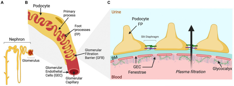
The glomerulus is a compact cluster of capillaries responsible for blood filtration and initiating urine production in the renal nephrons. A trilaminar structure in the capillary wall forms the glomerular filtration barrier (GFB), composed of glycocalyx-enriched and fenestrated endothelial cells adhering to the glomerular basement membrane and specialized visceral epithelial cells, podocytes, forming the outermost layer with a molecular slit diaphragm between their interdigitating foot processes. The unique dynamic and selective nature of blood filtration to produce urine requires the functionality of each of the GFB components, and hence, mimicking the glomerular filter in vitro has been challenging, though critical for various research applications and drug screening. Research efforts in the past few years have transformed our understanding of the structure and multifaceted roles of the cells and their intricate crosstalk in development and disease pathogenesis. In this review, we present a new wave of technologies that include glomerulus-on-a-chip, three-dimensional microfluidic models, and organoids all promising to improve our understanding of glomerular biology and to enable the development of GFB-targeted therapies. Here, we also outline the challenges and the opportunities of these emerging biomimetic systems that aim to recapitulate the complex glomerular filter, and the evolving perspectives on the sophisticated repertoire of cellular signaling that comprise the glomerular milieu.

Keywords: 3D model; crosstalk; glomerular endothelial cell; glomerular filtration barrier; in vitro; podocyte.


Conflict of interest statement

The authors declare that the research was conducted in the absence of any commercial or financial relationships that could be construed as a potential conflict of interest.

Figures

Figure 1
Figure 1
Schematic drawing of a single nephron and glomerulus, a glomerular capillary vessel, and the glomerular filtration barrier (GFB). (A) A single nephron comprising the blood-filtering glomerulus, enveloped by Bowman’s capsule that connects to the proximal tubule at the start of the urine-modifying tubular system. (B) The luminal surface of the glomerular capillary vessel is covered by glomerular endothelial cells (GECs), while podocytes wrap around the outside of the vessel with primary and foot processes (FT), forming an interdigitated pattern. Neighboring FPs are bridged by the slit diaphragm, one of the several essential components for glomerular permselectivity. The blood is filtered over this capillary barrier, and the pre-urine produced is forwarded from the Bowman’s capsule into the lumen of the proximal tubule. (C) The schematic cross section of the GFB displays the GEC fenestrae attached to the basement membrane (BM) and covered luminally by glycoproteins, proteoglycans, and glycosaminoglycans of the glycocalyx, important for maintaining the charge selectivity of the GFB. On the opposite side of the BM, the podocyte FPs are attached. The FPs interlink by slit diaphragm proteins, such as nephrin and podocin, are important for the restriction of albumin by the GFB. The arrow shows the direction of plasma filtration over the barrier.

Similar articles

Cited by

References

    1. Abe H., Sakurai A., Ochi A. (2019). Induction of steady-state glomeruloid sphere by self-assembly from human embryonic kidney cells. Biochem. Biophys. Res. Commun. 508, 654–659. 10.1016/j.bbrc.2018.11.160, PMID: - DOI - PubMed
    1. Agarwal A., Goss J. A., Cho A., McCain M. L., Parker K. K. (2013). Microfluidic heart on a chip for higher throughput pharmacological studies. Lab Chip 13, 3599–3608. 10.1039/c3lc50350j, PMID: - DOI - PMC - PubMed
    1. Anandakrishnan N., Azeloglu E. U. (2020). Kidney tissue engineering for precision medicine. Nat. Rev. Nephrol. 16, 623–624. 10.1038/s41581-020-00355-6, PMID: - DOI - PubMed
    1. Azeloglu E. U., Hardy S. V., Eungdamrong N. J., Chen Y., Jayaraman G., Chuang P. Y., et al. . (2014). Interconnected network motifs control podocyte morphology and kidney function. Sci. Signal. 7:ra12. 10.1126/scisignal.2004621, PMID: - DOI - PMC - PubMed
    1. Ballermann B. J. (2007). Contribution of the endothelium to the glomerular permselectivity barrier in health and disease. Nephron Physiol. 106, 19–25. 10.1159/000101796, PMID: - DOI - PubMed