Review
. 2021 Jun 4:12:676723.
doi: 10.3389/fneur.2021.676723. eCollection 2021.

Translations of Steinhausen's Publications Provide Insight Into Their Contributions to Peripheral Vestibular Neuroscience

Affiliations
Review

Translations of Steinhausen's Publications Provide Insight Into Their Contributions to Peripheral Vestibular Neuroscience

Hans Straka et al. Front Neurol. .

Abstract

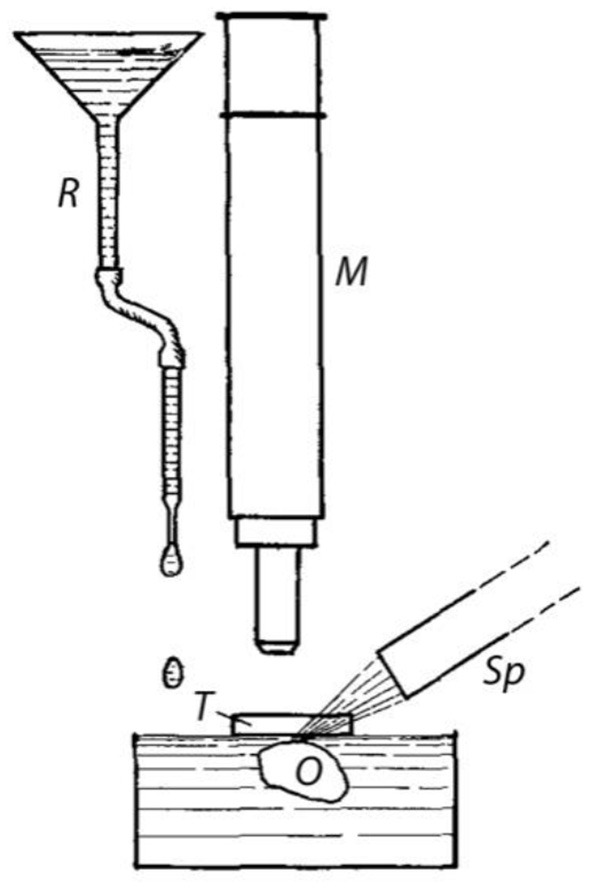
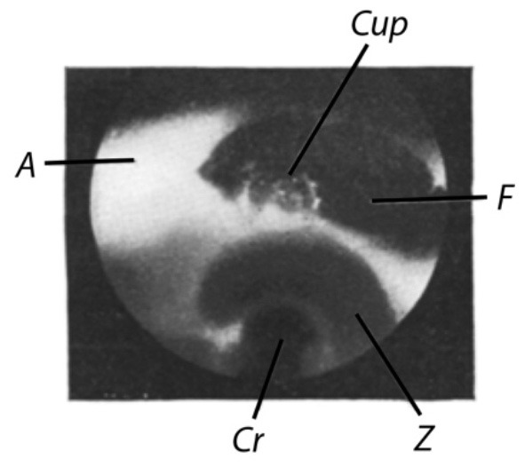
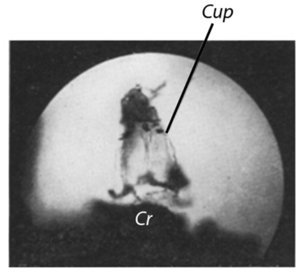
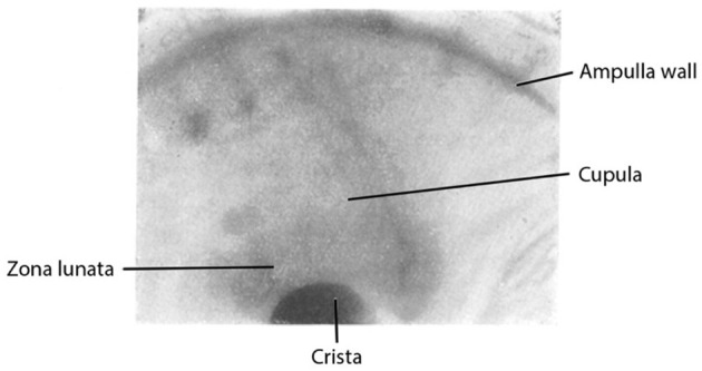
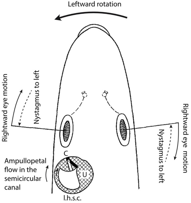
The quantitative relationship between angular head movement and semicircular canal function is most often referenced to the well-known torsion-pendulum model that predicts cupular displacement from input head acceleration. The foundation of this model can be traced back to Steinhausen's series of papers between 1927 and 1933 whereby he endeavored to document observations of cupular displacements that would directly infer movement of the endolymph resulting from angular rotation. He also was the first to establish the direct relationship between cupular displacement and compensatory eye movements. While the chronology of these findings, with their successes and pitfalls, are documented in Steinhausen's work, it reflects a fascinating journey that has been inaccessible to the non-German speaking community. Therefore, the present compilation of translations, with accompanying introduction and discussion, was undertaken to allow a larger component of the vestibular scientific community to gain insight into peripheral labyrinthine mechanics provided by this historical account.

Keywords: biomechanical model; crista; cupula; endolymph; labyrinth; torsion-pendulum.


Figures

Figure 1
Figure 1
(A) Ampulla of the anterior semicircular canal of the pike with incised window in which the cupula (Cup) can be seen. R = edges of the window. Freshly-produced preparation. The dark areas are Chinese ink particles. Lateral slit lamp illumination. Image captured on May 30, 1927 at 5.30. Beginning of the dissection at 2.10. Exposure time 9 min (to obtain the best possible depth effect, the exposure was set to 3 min, respectively, at 3 different depths). Objective Leitz 3. Ocular Leitz 1. Magnification 50x (Image number 30) (B). Same preparation. Cupula in motion (cf. text). Captured at 5.00, otherwise as (A) (Image number 29).
Figure 2
Figure 2
Device for the generation of fluid flow. O = Object, i.e., preparation with opened ampulla; T = Slide as protection against image distortion (cf. text); M = microscope; Sp = slit lamp; R = reservoir and tube for applying drops onto the water surface.
Figure 3
Figure 3
Cupula of a pike. Fixed preparation. Pike with a weight of about 0.5 kg. Dissected on May 14, 1927. Image captured on May 18, 1927. Lateral slit lamp illumination. Exposure time: 4 min. Cup = cupula; A = ampulla; F = window; Z = zona lunata; Cr = crista (Image number 10).
Figure 4
Figure 4
(A) Cupula of a pike. Isolated ampulla of the horizontal semicircular canal. Fixed preparation. The ampullary wall is completely ablated except for vestiges of the zona lunata (Z). Pike with a weight of about 2.25 kg. Dissected on March 8, 1927, image captured on May 20, 1927. Illumination: transmitted light. Exposure time: 1 min. Objective: Leitz 3, Ocular: Leitz 8. Magnification 80x (Image number 16) (B). Same preparation as in (A). Cupula in motion [cf. text for (B)]. All other as in (A) (Image number 17).
Figure 5
Figure 5
Cupula of a pike. From the posterior vertical ampulla. Pike with a weight of 0.5 kg. Fixed preparation. Dissected on May 20, 1927, image captured on May 24, 1927. Transmitted light. Exposure time 1 min. Objective L3, Ocular L3. Magnification 80x (Image number 18).
Figure 1
Figure 1
Stereo image of an ink-stained cupula in the intact semicircular canal ampulla of a freshly-produced pike labyrinth [Note by the translators: the two labels indicated by *(i.e., Ampulla*and Barberry spine*) were erroneously placed in the original paper and should be swapped].
Figure 2
Figure 2
Ink-stained freshly-produced cupula in the intact semicircular canal ampulla of the labyrinth of a pike. Magnification about 25x.
Figure 3
Figure 3
Ink-stained cupula in the intact semicircular canal ampulla of the labyrinth of a pike (freshly-produced) at rest (the 4 edges of the cupula are particularly clearly stained).
Figure 4
Figure 4
Same preparation as in Figure 3. Caloric stimulation. Three different deflection positions are photographically superimposed on a stationary plate (see also text).
Figure 1
Figure 1
Head of a pike viewed from dorsal with delineated left horizontal semicircular canal and utricle (schematic).
Figure 2
Figure 2
Semicircular canal with ampulla and cupula (schematic).
Figure 3
Figure 3
Cupula movement during long rotation periods. Abscissa: time (scale marks 30 = 40 seconds); ordinate, cupula movement as arbitrary scale.
Figure 4
Figure 4
Cupula movement during short rotation periods. Descriptions as in Figure 3.

References

    1. Van de Water TR. Historical aspects of inner ear anatomy and biology that undelie the design of hearing and balance prosthetic devices. Anatomical Rec. (2012) 295:1741–59. 10.1002/ar.22598 - DOI - PubMed
    1. Canalis RF, Mira E, Bonandrini L, Hinojosa R. Antonio Scarpa and the discovery of the membranous inner ear. Otol Neurotol. (2001) 22:105–12. 10.1097/00129492-200101000-00020 - DOI - PubMed
    1. Breuer J. Über die function der bogengänge des ohrlabyrinths. Med Jahrbücher. (1874) 4:72–124.
    1. Retzius G. Das Gehörorgan der Wirbelthiere. Stockholm: Samson & Wallin; (1881).
    1. Bechterew W. Ergebnisse der Durchschneidung des N. acusticus, nebst Erörterung der Bedeutung der semicirculären Canäle für das Körpergleichgewicht. Archiv Gesamte Phys Menschen Tiere. (1883) 30:312–47. 10.1007/BF01674334 - DOI

LinkOut - more resources