. 2021 Jun 2:12:665208.
doi: 10.3389/fimmu.2021.665208. eCollection 2021.

Rodent Models of Spondyloarthritis Have Decreased White and Bone Marrow Adipose Tissue Depots

Affiliations

Rodent Models of Spondyloarthritis Have Decreased White and Bone Marrow Adipose Tissue Depots

Giulia Furesi et al. Front Immunol. .

Abstract

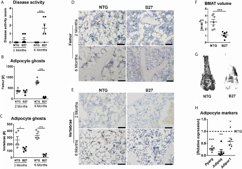
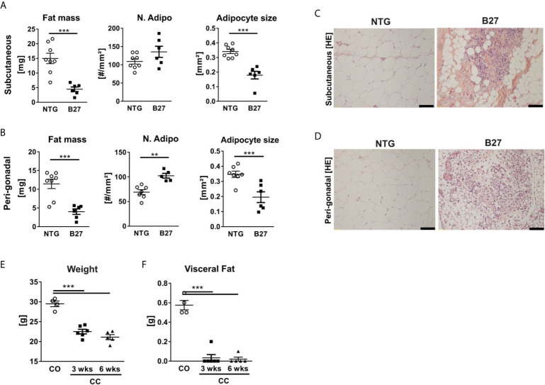
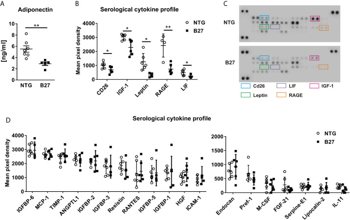
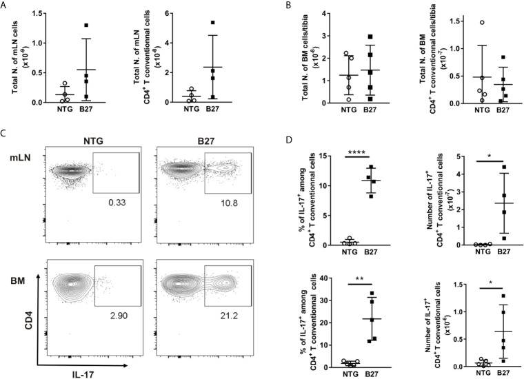
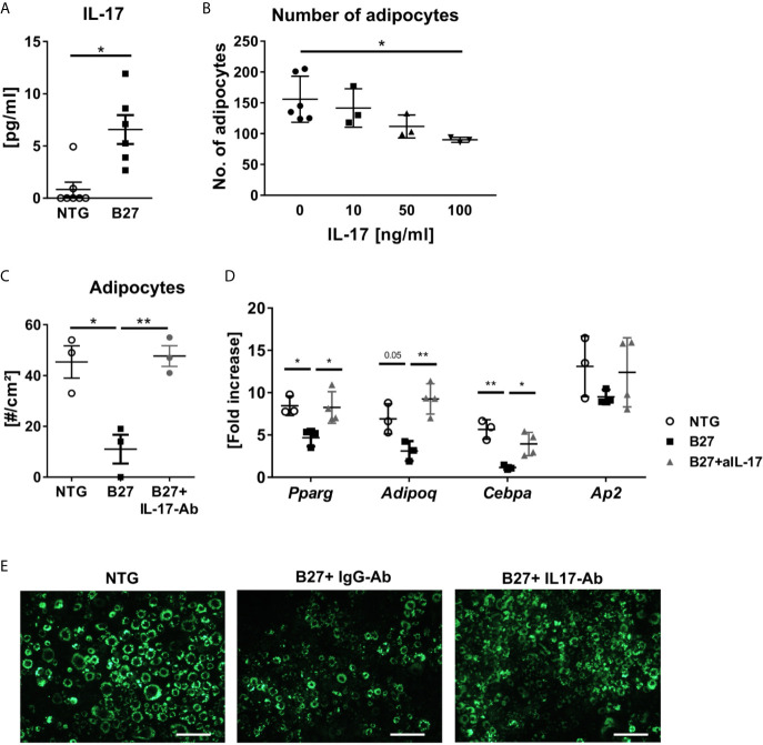
Bone marrow adipose tissue (BMAT) has recently been recognized as a distinct fat depot with endocrine functions. However, if and how it is regulated by chronic inflammation remains unknown. Here, we investigate the amount of white fat and BMAT in HLA-B27 transgenic rats and curdlan-challenged SKG mice, two well-established models of chronic inflammatory spondyloarthritis (SpA). Subcutaneous and gonadal white adipose tissue and BMAT was reduced by 65-70% and by up to 90% in both experimental models. Consistently, B27 rats had a 2-3-fold decrease in the serum concentrations of the adipocyte-derived cytokines adiponectin and leptin as well as a 2-fold lower concentration of triglycerides. The bone marrow of B27 rats was further characterized by higher numbers of neutrophils, lower numbers of erythroblast precursors, and higher numbers of IL-17 producing CD4+ T cells. IL-17 concentration was also increased in the serum of B27 rats. Using a cell culture model, we show that high levels of IL-17 in the serum of B27 rats negatively impacted adipogenesis (-76%), an effect that was reversed in the presence of neutralizing anti-IL-17 antibody. In summary, these findings show BMAT is severely reduced in two experimental models of chronic inflammatory SpA and suggest that IL-17 is involved in this process.

Keywords: HLA-B27 transgenic rat; IL-17; SKG mouse; bone marrow fat; spondyloarthritis.


Conflict of interest statement

The authors declare that the research was conducted in the absence of any commercial or financial relationships that could be construed as a potential conflict of interest.

Figures

Figure 1
Figure 1
Decreased numbers of bone marrow adipocytes in B27 transgenic rats. (A) Clinical score of 2 and 6 months old NTG and B27 rats. Disease level was graded from 0-3: 0=no inflammation, 1=arthritis on one leg, 2=arthritis on both legs, 3=severe arthritis, and other organ manifestation. (B) Number of bone marrow adipocytes ghosts from the femur of 2- and 6 months old NTG and B27 (n=5-6 per group). (C) Number of bone marrow adipocytes ghosts from the vertebrae of 2- and 6 months old NTG and B27 (n=5 per group). (D, E) Representative histology sections from femur and vertebrae of 2-and 6 months old NTG and B27 rats stained with von Kossa/toluidine blue (scale bar: 100 µm) (F, G) Osmium tetroxide staining for bone marrow adipose tissue (BMAT) followed by micro-computed tomography (μCT) analysis. Quantification of fat volume (F) and representative images (G) of stained lipids in femora of NTG and B27 rats (n=6-7 per group). (H) Relative expression of PPARγ, adiponectin, and adiponectin receptor in the bone marrow of NTG and B27 rats. Results are presented as individual dots with the mean indicated as horizontal line (n=5-6 per group). *p < 0.05; **p < 0.01 and ***p ≤ 0.001 vs. controls via unpaired Student’s t-test.
Figure 2
Figure 2
Decreased numbers of bone marrow adipocytes in SKG mice. (A, B) Clinical score of SKG animals at steady-state (SS) or curdlan-challenged (CC) for 3 and 6 weeks. (C) Osmium tetroxide staining for BMAT followed by μCT analysis. Quantification of fat volume (left) and representative images (right) of stained lipids in femora of SKG mice at 3- and 6-weeks post-curdlan challenge vs. control group. (D) Number of bone marrow adipocytes ghosts (left) and representative histology sections (right) from femur in SKG mice at 3 and 6 weeks post-curdlan challenge vs. control group stained with von Kossa/toluidine blue (scale bar: 100 µm). Results are presented as individual dots with the mean indicated as horizontal line (n=4-6 per group). *p < 0.05, **p ≤ 0.01, and ***p ≤ 0.001 vs. controls via two-way ANOVA with Tukey’s post hoc test (A, B) and one-way ANOVA with Tukey’s post hoc test (C, D).
Figure 3
Figure 3
Chronic inflammation in rodent models of SpA leads to loss of white adipose tissue. (A, B) Fat mass, number, and size of adipocytes in subcutaneous (top) and peri-gonadal (bottom) adipose tissue of NTG and B27 rats. (C, D) Representative histology sections stained with hematoxylin/eosin staining. Scale bars: 100 μm (n=6-8 per group). (E, F) Bodyweight and visceral fat weight of SKG mice at 3- and 6-weeks post-curdlan challenge vs. control group (n=4-6 per group). Results are presented as individual dots with the mean indicated as horizontal line. **p ≤ 0.01, and ***p ≤ 0.001 vs controls via unpaired Student´s t-test (A, B) and one-way ANOVA with Tukey’s post hoc test (E, F).
Figure 4
Figure 4
Altered metabolic profile in B27 transgenic rats. (A) Serum concentrations of adiponectin in NTG and B27 rats (n=5-7 per group). (B) Significantly regulated cytokines in the serum of NTG and B27 rats. (C) Representative image of cytokine profiler array results from serum of NTG and B27 rats. Significant ones are highlighted. (D) Relative levels of not-regulated obesity-related cytokines in NTG and B27 rats (B,D n=6-8 per group). Data are presented as mean ± SD. *p ≤ 0.05 and **p ≤ 0.001 NTG vs. B27 via unpaired Student’s t-test.
Figure 5
Figure 5
Expansion of IL17+ producing CD4+ T cells in the bone marrow from B27 transgenic rats. (A) Absolute number of live cells and CD4+ T cells in mesenteric lymph nodes (mLN) from NTG (grey) and B27 (white) rats were determined using flow cytometry. (B) Absolute number of live cells and CD4+ T cells in bone marrow (BM) from NTG and B27 rats. (C) Representative plots of intracellular IL-17 staining gated on CD4+ Foxp3- T cells in mLN (top) and BM (bottom) from NTG (left panel) or B27 (right panel) rats. (D) Frequencies of IL-17+ cells among CD4+ T cells (left) and their absolute numbers (right) in mLN (top) and BM (bottom) from NTG and B27 rats (A-D n=4-5 per group). Results are presented as individual dots with the mean indicated as horizontal line. Data were analyzed by unpaired Student’s t-test (*p<0.05, **p < 0.01 and ****p < 0.0001).
Figure 6
Figure 6
IL-17 from B27 transgenic rat serum inhibits adipogenesis in vitro. (A) IL-17 levels in sera of in B27 and NTG rats (n=6-7 per group). (B) Absolute numbers of 3T3 adipocytes after exposure to control medium or increasing doses of IL-17 during adipocyte differentiation (n=3-6 per group). (C, D) Absolute numbers of 3T3 adipocytes and relative expression of the most representative adipocyte markers after 48 h exposure to 5% serum from NTG rats, B27 rats + IgG antibody (IgG-Ab), or B27 rats + neutralizing anti-IL-17 antibody (IL-17-Ab) treatment (n=3-6 per group). (E) Representative fluorescence microscopy pictures of LipidTOX stained 3T3 cells. Scale bar: 50 µm. All experiments were performed independently three times. Results are presented as individual dots with the mean indicated as horizontal line. *p ≤ 0.05 and **p ≤ 0.01 NTG vs. B27 via unpaired Student’s t-test (A). and one-way ANOVA with Tukey’s post hoc test (B–D).

Similar articles

Cited by

References

    1. Suchacki KJ, Tavares AAS, Mattiucci D, Scheller EL, Papanastasiou G, Gray C, et al. . Bone Marrow Adipose Tissue Is a Unique Adipose Subtype With Distinct Roles in Glucose Homeostasis. Nat Commun (2020) 11:3097. 10.1038/s41467-020-16878-2 - DOI - PMC - PubMed
    1. Scheller EL, Cawthorn WP, Burr AA, Horowitz MC, MacDougald OA. Marrow Adipose Tissue: Trimming the Fat. Trends Endocrinol Metab TEM (2016) 27:392–403. 10.1016/j.tem.2016.03.016 - DOI - PMC - PubMed
    1. Sulston RJ, Learman BS, Zhang B, Scheller EL, Parlee SD, Simon BR, et al. . Increased Circulating Adiponectin in Response to Thiazolidinediones: Investigating the Role of Bone Marrow Adipose Tissue. Front Endocrinol (2016) 7:128. 10.3389/fendo.2016.00128 - DOI - PMC - PubMed
    1. Krings A, Rahman S, Huang S, Lu Y, Czernik PJ, Lecka-Czernik B. Bone Marrow Fat Has Brown Adipose Tissue Characteristics, Which Are Attenuated With Aging and Diabetes. Bone (2012) 50:546–52. 10.1016/j.bone.2011.06.016 - DOI - PMC - PubMed
    1. Chen Q, Shou P, Zheng C, Jiang M, Cao G, Yang Q, et al. . Fate Decision of Mesenchymal Stem Cells: Adipocytes or Osteoblasts? Cell Death Differ (2016) 23:1128–39. 10.1038/cdd.2015.168 - DOI - PMC - PubMed

Publication types

LinkOut - more resources