. 2021 Jun 2:12:680838.
doi: 10.3389/fgene.2021.680838. eCollection 2021.

Oligogenic Inheritance of Monoallelic TRIP11, FKBP10, NEK1, TBX5, and NBAS Variants Leading to a Phenotype Similar to Odontochondrodysplasia

Affiliations

Oligogenic Inheritance of Monoallelic TRIP11, FKBP10, NEK1, TBX5, and NBAS Variants Leading to a Phenotype Similar to Odontochondrodysplasia

Alice Costantini et al. Front Genet. .

Abstract

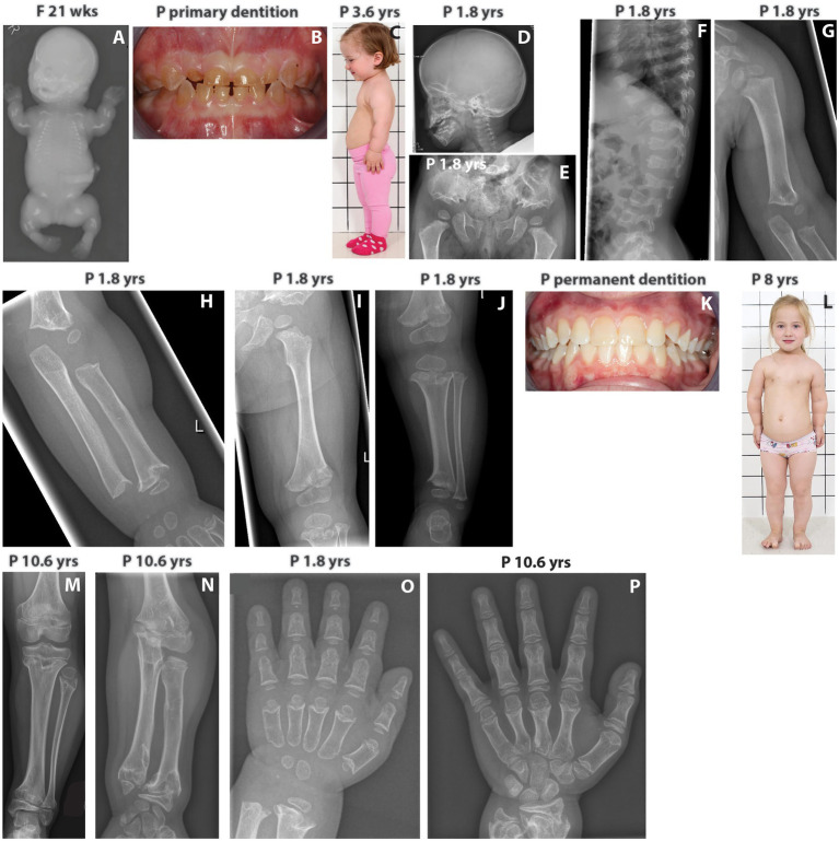
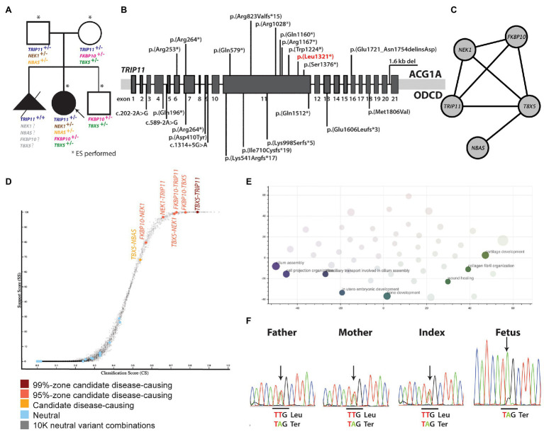
Skeletal dysplasias are often well characterized, and only a minority of the cases remain unsolved after a thorough analysis of pathogenic variants in over 400 genes that are presently known to cause monogenic skeletal diseases. Here, we describe an 11-year-old Finnish girl, born to unrelated healthy parents, who had severe short stature and a phenotype similar to odontochondrodysplasia (ODCD), a monogenic skeletal dysplasia caused by biallelic TRIP11 variants. The family had previously lost a fetus due to severe skeletal dysplasia. Exome sequencing and bioinformatic analysis revealed an oligogenic inheritance of a heterozygous nonsense mutation in TRIP11 and four likely pathogenic missense variants in FKBP10, TBX5, NEK1, and NBAS in the index patient. Interestingly, all these genes except TBX5 are known to cause skeletal dysplasia in an autosomal recessive manner. In contrast, the fetus was found homozygous for the TRIP11 mutation, and achondrogenesis type IA diagnosis was, thus, molecularly confirmed, indicating two different skeletal dysplasia forms in the family. To the best of our knowledge, this is the first report of an oligogenic inheritance model of a skeletal dysplasia in a Finnish family. Our findings may have implications for genetic counseling and for understanding the yet unsolved cases of rare skeletal dysplasias.

Keywords: TRIP11; achondrogenesis type IA; odontochondrodysplasia; oligogenic inheritance; short stature.


Conflict of interest statement

The authors declare that the research was conducted in the absence of any commercial or financial relationships that could be construed as a potential conflict of interest.

Figures

Figure 1
Figure 1
(A) Babygram of the deceased fetus shows extremely short tubular bones, very narrow thorax, horizontally oriented short ribs, and retarded ossification of the vertebrae. (B) Patient’s primary dentition showing dentinogenesis imperfecta. (C) The index patient at 3.6 yrs has short neck and limbs, redundant skin folds in the arms, and protuberant abdomen. (D) The skull of the index shows mild frontal bossing. (E) Patient’s pelvis showing trident configuration of the acetubulum and lacy iliac wings as well as short femoral necks with metaphyseal irregularities. (F) Patient’s spine displaying abnormal vertebrae with coronal clefts. (G–I) X-rays showing short and broad humerus with metaphyseal changes and shortening of the tibia and ulna as well as flared metaphyses. (J) The lower limb of the index patient displays short long bones, short femoral neck, marked metaphyseal irregularities, and normal epiphyses. (K) Patient’s permanent dentition with no signs of dentinogenesis imperfecta. (L) The index at the age of 8 yrs has normal facial features, short neck, redundant skin folds in the arms, narrow chest, prominent sternum, short limbs, mesomelic shortening of the upper arm and brachydactyly. (M) At 10.6 yrs, the index patient has marked metaphyseal irregularities of the tibia and ulna. (N) At 10.6 yrs, overgrowth of the fibula is noticed. (O,P) Hands of the index at 1.8 and 10.6 yrs. Brachydactyly and distinctive changes with deeper cupping of the metaphyses are evident. Progressive metaphyseal changes of the distal radius and ulna are also seen at 10.6 yrs. F, fetus; P, index patient; wks, weeks; yrs, years.
Figure 2
Figure 2
(A) Pedigree of the family and rare mutations identified in the genes presently linked to skeletal dysplasia. (B) Biallelic TRIP11 mutations that are presently known to cause ACG1A and ODCD. The mutation that has also been identified in the present study is marked with red. (C) Gene network showing the interaction between the genes in which rare mutations have been identified. (D) Genes affected by variants suggesting digenic inheritance. (E) Biological processes in which the genes suggesting oligogenic inheritance are involved. (F) Sanger sequencing of the TRIP11 region affected by the nonsense p.(Leu1321*) mutation. ACG1A, achondrogenesis type IA; ODCD, odontochondrodysplasia.

References

    1. Alanay Y., Avaygan H., Camacho N., Utine G. E., Boduroglu K., Aktas D., et al. . (2010). Mutations in the gene encoding the RER protein FKBP65 cause autosomal-recessive osteogenesis imperfecta. Am. J. Hum. Genet. 86, 551–559. 10.1016/j.ajhg.2010.02.022, PMID: - DOI - PMC - PubMed
    1. Ameur A., Dahlberg J., Olason P., Vezzi F., Karlsson R., Martin M., et al. . (2017). SweGen: a whole-genome data resource of genetic variability in a cross-section of the Swedish population. Eur. J. Hum. Genet. 25, 1253–1260. 10.1038/ejhg.2017.130, PMID: - DOI - PMC - PubMed
    1. Aoki T., Ichimura S., Itoh A., Kuramoto M., Shinkawa T., Isobe T., et al. . (2009). Identification of the neuroblastoma-amplified gene product as a component of the syntaxin 18 complex implicated in Golgi-to-endoplasmic reticulum retrograde transport. Mol. Biol. Cell 20, 2639–2649. 10.1091/mbc.e08-11-1104, PMID: - DOI - PMC - PubMed
    1. Barnes A. M., Cabral W. A., Weis M., Makareeva E., Mertz E. L., Leikin S., et al. . (2012). Absence of FKBP10 in recessive type XI osteogenesis imperfecta leads to diminished collagen cross-linking and reduced collagen deposition in extracellular matrix. Hum. Mutat. 33, 1589–1598. 10.1002/humu.22139, PMID: - DOI - PMC - PubMed
    1. Chen Y., Chen C. F., Riley D. J., Chen P. L. (2011). Nek1 kinase functions in DNA damage response and checkpoint control through a pathway independent of ATM and ATR. Cell Cycle 10, 655–663. 10.4161/cc.10.4.14814, PMID: - DOI - PMC - PubMed

LinkOut - more resources