

Y-shaped Muscular Wrapping Technique Avoiding Re-infection of a Replaced Aortic Graft: A Cadaveric Study
- PMID: 34150424
- PMCID: PMC8208446
- DOI: 10.1097/GOX.0000000000003626
Y-shaped Muscular Wrapping Technique Avoiding Re-infection of a Replaced Aortic Graft: A Cadaveric Study
Abstract
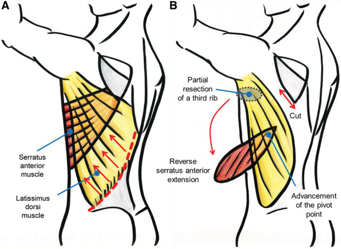
Replacing an infected prosthetic thoracic aorta graft carries a high re-infection risk. We previously reported two clinical cases successfully treated with a new muscular wrapping technique: latissimus dorsi (LD) muscle flap with a distally based serratus anterior (SA) extension; however, a cadaveric study to prove the regular existence of the distal attachment area was lacking. We tried to establish an appropriate way of elevating the combined muscle flap safely. All of the cadavers were preserved using the Thiel embalming technique to retain flexibility. We checked for the existence of the distal attachment area between the LD and SA. Combined muscle flaps were elevated proximally while identifying the thoracodorsal artery, including the LD and SA branches. After the SA branch was ligated and cut, the SA muscle was manually peeled from the LD muscle with only the distal tight attachment area remaining. Contrast-enhanced computed tomography was performed using a multislice computed tomography system. Six human cadavers (three men, three women: 91 years old, on average) were examined. All six LD and SA combined muscle flaps showed a distal tight attachment area at the level from the seventh rib to the ninth rib. The tip of the SA muscle easily reached the sternum. Contrast-enhanced computed tomography failed to reconfirm the distal vascular flow from the LD to the reverse SA muscle, which we had visualized in a clinical case. We demonstrated the anatomical reliability of the new Y-shaped muscular flaps, which are suitable for preventing re-infection of aortic graft replacement.
Copyright © 2021 The Authors. Published by Wolters Kluwer Health, Inc. on behalf of The American Society of Plastic Surgeons.
Conflict of interest statement
Disclosure: All the authors have no financial interest to declare in relation to the content of this article.
Figures


Similar articles
-
Planned Y-shaped Muscle Wrapping for Salvaging Aortic Graft Infection: Latissimus Dorsi and Reverse Serratus Anterior Muscles.Plast Reconstr Surg Glob Open. 2024 Nov 25;12(11):e6350. doi: 10.1097/GOX.0000000000006350. eCollection 2024 Nov. Plast Reconstr Surg Glob Open. 2024. PMID: 39600326 Free PMC article.
-
The versatility of the thoracodorsal artery based composite flaps with vascularized rib and a systematic review of the literature.J Surg Oncol. 2019 Sep;120(3):527-539. doi: 10.1002/jso.25579. Epub 2019 Jun 13. J Surg Oncol. 2019. PMID: 31197840
-
One-stage free transfer of latissimus dorsi-serratus anterior combined muscle flap with dual innervation for smile reanimation in established facial paralysis.J Plast Reconstr Aesthet Surg. 2020 Jun;73(6):1107-1115. doi: 10.1016/j.bjps.2020.01.032. Epub 2020 Jan 31. J Plast Reconstr Aesthet Surg. 2020. PMID: 32334999
-
A comparative evaluation of intrathoracic latissimus dorsi and serratus anterior muscle transposition.Eur J Cardiothorac Surg. 2000 Oct;18(4):435-9. doi: 10.1016/s1010-7940(00)00538-8. Eur J Cardiothorac Surg. 2000. PMID: 11024381
-
Anatomical bases of the bypass-flap: study of the thoracodorsal axis.Surg Radiol Anat. 2005 Apr;27(2):86-93. doi: 10.1007/s00276-004-0299-y. Epub 2005 Jan 19. Surg Radiol Anat. 2005. PMID: 15657635
References
-
- Luehr M, Etz CD, Nozdrzykowski M, et al. . Emergency open surgery for aorto-oesophageal and aorto-bronchial fistulae after thoracic endovascular aortic repair: a single-centre experience. Eur J Cardiothorac Surg. 2015; 47:374–82; discussion 382. - PubMed
-
- Takano T, Terasaki T, Wada Y, et al. . Treatment of prosthetic graft infection after thoracic aorta replacement. Ann Thorac Cardiovasc Surg. 2014; 20:304–309. - PubMed
-
- Thiel W. [The preservation of the whole corpse with natural color]. Ann Anat. 1992; 174:185–195. - PubMed
-
- Frautschi RS, Bassiri Gharb B, Duong MM, et al. . The cardioplastic approach to the treatment of infected aortic grafts. Ann Plast Surg. 2017; 79:221–225. - PubMed
LinkOut - more resources
Full Text Sources
Research Materials