Review
. 2021 Aug;11(8):2344-2361.
doi: 10.1016/j.apsb.2021.02.010. Epub 2021 Feb 16.

Microneedle-based devices for point-of-care infectious disease diagnostics

Affiliations
Review

Microneedle-based devices for point-of-care infectious disease diagnostics

Rachael V Dixon et al. Acta Pharm Sin B. 2021 Aug.

Abstract

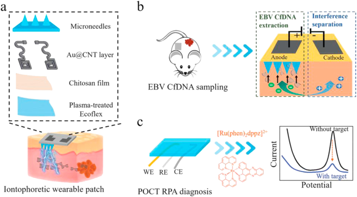
Recent infectious disease outbreaks, such as COVID-19 and Ebola, have highlighted the need for rapid and accurate diagnosis to initiate treatment and curb transmission. Successful diagnostic strategies critically depend on the efficiency of biological sampling and timely analysis. However, current diagnostic techniques are invasive/intrusive and present a severe bottleneck by requiring specialist equipment and trained personnel. Moreover, centralised test facilities are poorly accessible and the requirement to travel may increase disease transmission. Self-administrable, point-of-care (PoC) microneedle diagnostic devices could provide a viable solution to these problems. These miniature needle arrays can detect biomarkers in/from the skin in a minimally invasive manner to provide (near-) real-time diagnosis. Few microneedle devices have been developed specifically for infectious disease diagnosis, though similar technologies are well established in other fields and generally adaptable for infectious disease diagnosis. These include microneedles for biofluid extraction, microneedle sensors and analyte-capturing microneedles, or combinations thereof. Analyte sampling/detection from both blood and dermal interstitial fluid is possible. These technologies are in their early stages of development for infectious disease diagnostics, and there is a vast scope for further development. In this review, we discuss the utility and future outlook of these microneedle technologies in infectious disease diagnosis.

Keywords: AC, alternating current; APCs, antigen-presenting cells; ASSURED, affordable, sensitive, specific, user-friendly, rapid and robust, equipment-free and deliverable to end-users; Biomarker detection; Biosensor; CMOS, complementary metal-oxide semiconductor; COVID, coronavirus disease; COVID-19; CSF, cerebrospinal fluid; CT, computerised tomography; CV, cyclic voltammetry; DC, direct current; DNA, deoxyribonucleic acid; DPV, differential pulse voltammetry; EBV, Epstein–Barr virus; EDC/NHS, 1-ethyl-3-(3-dimethylaminoproply) carbodiimide/N-hydroxysuccinimide; ELISA, enzyme-linked immunosorbent assay; GOx, glucose oxidase; HIV, human immunodeficiency virus; HPLC, high performance liquid chromatography; HRP, horseradish peroxidase; IP, iontophoresis; ISF, interstitial fluid; IgG, immunoglobulin G; Infectious disease; JEV, Japanese encephalitis virus; MN, microneedle; Microneedle; NA, nucleic acid; OBMT, one-touch-activated blood multidiagnostic tool; OPD, o-phenylenediamine; PCB, printed circuit board; PCR, polymerase chain reaction; PDMS, polydimethylsiloxane; PEDOT, poly(3,4-ethylenedioxythiophene); PNA, peptide nucleic acid; PP, polyphenol; PPD, poly(o-phenylenediamine); PoC, point-of-care; Point-of-care diagnostics (PoC); SALT, skin-associated lymphoid tissue; SAM, self-assembled monolayer; SEM, scanning electron microscope; SERS, surface-enhanced Raman spectroscopy; SWV, square wave voltammetry; Skin; TB, tuberculosis; UV, ultraviolet; VEGF, vascular endothelial growth factor; WHO, World Health Organisation; cfDNA, cell-free deoxyribonucleic acid.


Conflict of interest statement

The authors have no conflicts of interest to declare.

Figures

Image 1
Graphical abstract
Figure 1
Figure 1
Current microneedle diagnostic platforms function based on biofluid extraction using hollow (A) or solid (B) microneedles, specific target analyte capture (C) and electrochemical sensing (D). Adapted with permission from Ref. and licenced under CC BY 4.0.
Figure 2
Figure 2
Venn diagram showing the desired infectious disease PoC diagnostic traits and the relationship between current microneedle (MN)-based platforms.
Figure 3
Figure 3
Schematic images depicting various designs of microneedle arrays interfacing microfluidic on-chip analysis chambers. (A)‒(C) Schematic of a one-touch-activated blood multidiagnostic tool (OBMT). (A) OBMT complete device design and structure. (B) OBMT paper-based multiplex sensor. (C) Operating principle of the OBMT device. (D) Schematic of silicon dioxide transdermal biosensor showing the front side and backside of the microneedle chip, the ‘stand-alone’ sensor section and the full integrated device. (E) Schematic of a continuous glucose monitoring hollow silicon microneedle-based device with glucose sensing chamber. Permissions: (A)‒(C) Reprinted with permissions from Ref. under copyright© 2015, The Royal Society of Chemistry; permission conveyed through Copyright Clearance Center, Inc. (D) Adapted from Ref. under copyright© 2015, Elsevier. (E) Reprinted from Ref. under copyright© 2014, SAGE.
Figure 4
Figure 4
Images of different design strategies for hollow microneedles. (A–C) Images of microneedles with bevelled tip angles of 90°, 45° and 15°. (D)‒(E) Scanning electron microscope (SEM) images showing an array and single ‘hypodermic needle’ microneedle design where the orifice is shifted 25 μm off centre. (F)‒(G) SEM images showing an array and single ‘snake fang’ microneedle design where the orifice is shifted 50 μm off centre resulting in the opening on the side of the microneedle. (H)‒(I) SEM images of tapered and straight microneedle designs with the orifice at the tip. Permissions: (A)‒(C) Reprinted from Ref. under copyright© 2013, Springer Nature. (D)‒(G) Reprinted from Ref. under copyright© 2004, Elsevier. (H)‒(I) Reprinted from Ref. under copyright© 2013, Elsevier.
Figure 5
Figure 5
Images of solid, porous and hydrogel microneedle designs. (A)‒(C) Biofluid extraction device comprising of a row of 9 solid microneedles in series with absorbent paper attached. (D)‒(E) Optical micrograph and SEM image of a single porous microneedle fabricated from polydimethylsiloxane (PDMS). (F) Gelatin methacryloyl hydrogel microneedle array showing appearance change after different durations of fluid uptake. Permissions: (A)‒(C) Reprinted from Ref. under copyright© 2019, John Wiley & Sons. (D)‒(E) Reprinted from Ref. under copyright© 2019, Springer Nature. (F) Reprinted from Ref. under copyright© 2020, John Wiley & Sons.
Figure 6
Figure 6
Modes of electrochemical sensing for the detection of bacteria. (A)‒(B) Examples of indirect bacterial detection using direct current (DC)-based techniques. Square wave voltammetry (SWV) plots current vs. potential and chronoamperometry plots current vs. time. (A) Sensing of cell-secreted metabolites via redox reactions. (B) Sensing of exogenous bacterial enzymatic electroactive by-products. (C)‒(D) Examples of direct bacterial detection using impedimetric or alternating current (AC)-based techniques. (C) Bacterial binding event causes a change in impedance due to reduced electron transfer activity of a mediator as a result of surface passivation. Signal is typically represented on a Nyquist plot. (D) Bacterial binding event is measured by a change in capacitance as a function of AC frequency. Reprinted from Ref. under copyright© 2020, American Chemical Society.
Figure 7
Figure 7
Known antibody immobilisation strategies for immunosensors. (A) Uncontrolled antibody adsorption. (B) Protein mediated antibody orientation and immobilisation. (C) 1-Ethyl-3-(3-dimethylaminoproply) carbodiimide/N-hydroxysuccinimide (EDC/NHS) coupling creating robust covalent linkage via amine bond formation. (D) Reduction of antibody disulfides to create thiol groups for immobilisation onto gold surfaces. (E) Reduction of antibody disulfides for site specific coupling. (F) Oxidation of sugar chains to create reactive aldehyde groups. Image adapted with permission from Ref. and licenced under CC BY 4.0.
Figure 8
Figure 8
Paper-based detection of human TNF-alpha using analyte-capturing microneedles. Colour signals generated through the enzymatic reaction between OPD and HRP can be blotted on to paper, which concentrates the signal and offers information about the spatial distribution of the target biomarker. Reprinted from Ref. under copyright© 2015, Controlled Release Society.
Figure 9
Figure 9
Densitometric analysis used in conjunction with paper-based detection of cytokines from the skin, showing (A) multiplexing capabilities (IL-1alpha, IL-6 and assay controls) and signal visualisation, (B) signal quantification by densitometry, and (C) validation using standard plate-based ELISA. Reprinted from Ref. under copyright© 2015, Controlled Release Society.
Figure 10
Figure 10
Schematic depiction of an iontophoretic wearable and microfluidic electrochemical sensor based on microneedles: (A) construction of the microneedle device; (B) extraction of EBV cfDNA from the ISF in mice by reverse iontophoresis and entrapment in the hydrogel microneedles; (C) electrochemical quantification of the captured cfDNA using a 3-electrode system (WE: working electrode; RE: reference electrode; CE: counter electrode). Reprinted from Ref. under copyright © 2020, Wiley-VCH Verlag GmbH & Co. KGaA, Weinheim.

References

    1. Committee on Diagnostic Error in Health Care, Board on Health Care Services, Institute of Medicine . In: Improving diagnosis in health care. Balogh E.P., Miller B.T., Ball J.R., editors. National Academies Press (US); Washington, D.C.: 2015. The national academies of sciences, engineering, and medicine the diagnostic process. - PubMed
    1. Miró E.M., Sánchez N.P. In: Atlas of dermatology in internal medicine. Sánchez N.P., editor. Springer; New York, NY: 2012. Cutaneous manifestations of infectious diseases; pp. 77–119.
    1. Leptospirosis Information Centre Leptospirosis information - other infections with similar symptoms. Leptospirosis org. http://www.leptospirosis.org/other-infections-with-similar-symptoms/ Available from:
    1. Centers for disease control and prevention Epstein-Barr virus and infectious mononucleosis. https://www.cdc.gov/epstein-barr/about-ebv.html Available from:
    1. Paliwal S., Hwang B.H., Tsai K.Y., Mitragotri S. Diagnostic opportunities based on skin biomarkers. Eur J Pharmaceut Sci. 2013;50:546–556. - PubMed

LinkOut - more resources