

Evaluation of macular vessel density changes after vitrectomy with silicone oil tamponade in patients with rhegmatogenous retinal detachment
- PMID: 34150544
- PMCID: PMC8165621
- DOI: 10.18240/ijo.2021.06.14
Evaluation of macular vessel density changes after vitrectomy with silicone oil tamponade in patients with rhegmatogenous retinal detachment
Abstract
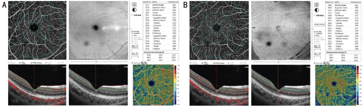
Aim: To evaluate macular microvasculature changes in eyes after pars plana vitrectomy (PPV) and intraocular silicone oil (SO) tamponade for macula-off rhegmatogenous retinal detachment (RRD) using optical coherence tomography angiography (OCTA).
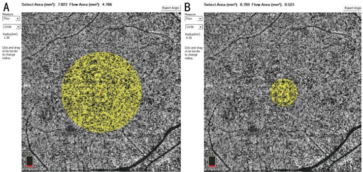
Methods: Totally 19 eyes (19 patients) with macula-off RRD who underwent PPV and intraocular SO tamponade were retrospectively reviewed. The parafoveal superficial capillary plexus (SCP) vessel density (VD), deep capillary plexus (DCP) VD, choriocapillaris plexus (CCP) VD, and foveal macular thickness were evaluated using OCTA throughout 16wk postoperatively. The values of healthy fellow eyes were used as control.
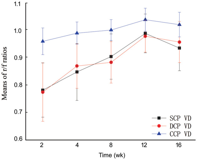
Results: The parafoveal SCP, DCP, and CCP VDs were significant increased over time in RRD eyes during the 12wk postoperatively, then decreased at 16wk postoperatively (all P<0.01). The ratios of RRD eyes and fellow healthy eyes (r/f ratios) of the SCP and DCP VDs were lower than those of the CCP VD postoperatively (all P<0.05). There were not significant differences in the r/f ratios between SCP and DCP VDs postoperatively (all P>0.05).
Conclusion: The parafoveal SCP, DCP, and CCP VDs gradually recover over time after PPV surgery with SO tamponade. Long-time SO tamponade might decrease postoperative macular VDs. Compared to parafoveal CCP VD, the parafoveal SCP and DCP VDs were more vulnerable in RRD eyes postoperatively.
Keywords: optical coherence tomography angiography; rhegmatogenous retinal detachment; silicone oil; vessel density; vitrectomy.
International Journal of Ophthalmology Press.
Figures
Similar articles
-
Effect of long-term silicone oil tamponade on the density of blood vessels in the macular and peripapillary region in patients with rhegmatogenous retinal detachment.Int Ophthalmol. 2025 Mar 25;45(1):124. doi: 10.1007/s10792-025-03460-2. Int Ophthalmol. 2025. PMID: 40131517
-
Macular microcirculation changes after macula-off rhegmatogenous retinal detachment repair with silicone oil tamponade evaluated by OCT-A: preliminary results.Ther Adv Ophthalmol. 2022 Jun 18;14:25158414221105222. doi: 10.1177/25158414221105222. eCollection 2022 Jan-Dec. Ther Adv Ophthalmol. 2022. PMID: 35734223 Free PMC article.
-
Microvascular changes on optical coherence tomography angiography after rhegmatogenous retinal detachment vitrectomy with silicone tamponade.PLoS One. 2021 Mar 12;16(3):e0248433. doi: 10.1371/journal.pone.0248433. eCollection 2021. PLoS One. 2021. PMID: 33711059 Free PMC article.
-
Microstructural and hemodynamic changes in the fundus after pars plana vitrectomy for different vitreoretinal diseases.Graefes Arch Clin Exp Ophthalmol. 2024 Jul;262(7):1977-1992. doi: 10.1007/s00417-023-06303-x. Epub 2023 Nov 20. Graefes Arch Clin Exp Ophthalmol. 2024. PMID: 37982887 Review.
-
Macular microcirculation changes after repair of rhegmatogenous retinal detachment assessed with optical coherence tomography angiography: A systematic review and meta-analysis.Front Physiol. 2022 Dec 14;13:995353. doi: 10.3389/fphys.2022.995353. eCollection 2022. Front Physiol. 2022. PMID: 36589420 Free PMC article.
Cited by
-
Long-Term Macular Vascular Changes after Primary Rhegmatogenous Retinal Detachment Surgery Resolved with Different Tamponade or Different Surgical Techniques.Life (Basel). 2022 Sep 30;12(10):1525. doi: 10.3390/life12101525. Life (Basel). 2022. PMID: 36294960 Free PMC article.
-
Retinal and Corneal Changes Associated with Intraocular Silicone Oil Tamponade.J Clin Med. 2022 Sep 5;11(17):5234. doi: 10.3390/jcm11175234. J Clin Med. 2022. PMID: 36079165 Free PMC article. Review.
-
Association between retinal vessel density and postoperative time after primary repair of rhegmatogenous retinal detachment.PLoS One. 2021 Oct 1;16(10):e0258126. doi: 10.1371/journal.pone.0258126. eCollection 2021. PLoS One. 2021. PMID: 34597349 Free PMC article.
-
Effect of silicone oil on retinal microcirculation after vitrectomy for rhegmatogenous retinal detachment evaluated by OCT angiography: a literature review.Ther Adv Ophthalmol. 2023 May 24;15:25158414231174145. doi: 10.1177/25158414231174145. eCollection 2023 Jan-Dec. Ther Adv Ophthalmol. 2023. PMID: 37255621 Free PMC article. Review.
-
Expression levels of ROS and Atg proteins in the vitreous in rhegmatogenous retinal detachment.Int J Ophthalmol. 2023 Mar 18;16(3):348-353. doi: 10.18240/ijo.2023.03.03. eCollection 2023. Int J Ophthalmol. 2023. PMID: 36935782 Free PMC article.
References
-
- Machemer R. The importance of fluid absorption, traction, intraocular currents, and chorioretinal scars in the therapy of rhegmatogenous retinal detachments. XLI Edward Jackson Memorial Lecture. Am J Ophthalmol. 1984;98(6):681–693. - PubMed
-
- van Bussel EM, van der Valk R, Bijlsma WR, La Heij EC. Impact of duration of macula-off retinal detachment on visual outcome: a systematic review and meta-analysis of literature. Retina. 2014;34(10):1917–1925. - PubMed
-
- Park DH, Choi KS, Sun HJ, Lee SJ. Factors associated with visual outcome after macula-off rhegmatogenous retinal detachment surgery. Retina. 2018;38(1):137–147. - PubMed
LinkOut - more resources
Full Text Sources
Miscellaneous