Review
. 2021 Jun 7:9:671402.
doi: 10.3389/fcell.2021.671402. eCollection 2021.

Organ-Specific Branching Morphogenesis

Affiliations
Review

Organ-Specific Branching Morphogenesis

Christine Lang et al. Front Cell Dev Biol. .

Abstract

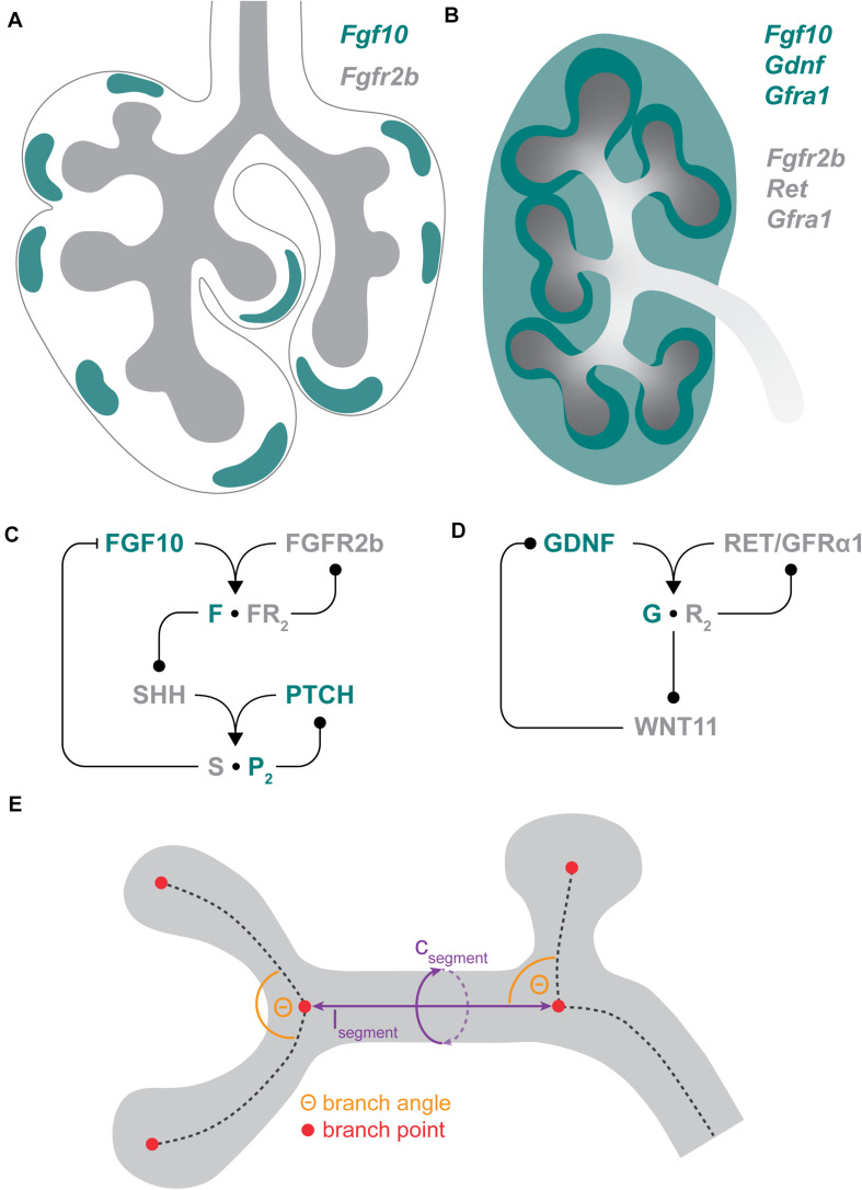
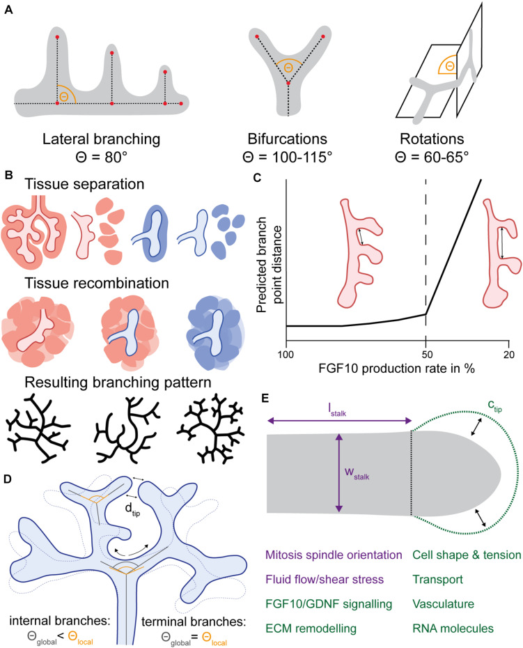
A common developmental process, called branching morphogenesis, generates the epithelial trees in a variety of organs, including the lungs, kidneys, and glands. How branching morphogenesis can create epithelial architectures of very different shapes and functions remains elusive. In this review, we compare branching morphogenesis and its regulation in lungs and kidneys and discuss the role of signaling pathways, the mesenchyme, the extracellular matrix, and the cytoskeleton as potential organ-specific determinants of branch position, orientation, and shape. Identifying the determinants of branch and organ shape and their adaptation in different organs may reveal how a highly conserved developmental process can be adapted to different structural and functional frameworks and should provide important insights into epithelial morphogenesis and developmental disorders.

Keywords: branch angle; branch distance; branch shape; branching morphogenesis; kidney; lung; tissue mechanics; turing pattern.


Conflict of interest statement

The authors declare that the research was conducted in the absence of any commercial or financial relationships that could be construed as a potential conflict of interest.

Figures

FIGURE 1
FIGURE 1
Branching morphogenesis. (A) Gene expression domains of Fgf10 and its receptor Fgfr2b in the E11.5 mouse lung. Fgf10 is expressed in a spotty pattern in the submesothelial mesenchyme (green), while Fgfr2b is expressed in the epithelium (gray). (B) Gene expression domains of Gdnf and Fgf10 and their receptors in the E12.5 murine kidney. Gdnf is expressed in both the cap mesenchyme (dark green) and the stroma (light green), while Fgf10 is expressed in the cap mesenchyme. Fgfr2b is expressed in the ureteric bud (UB), while Ret expression is restricted to UB tips (dark gray). The Ret co-receptor Gfrα1 is expressed both in the metanephric mesenchyme and the ureteric bud. (C) Core signaling network in lung branching morphogenesis. FGF10 signals via FGFR2b which enhances Shh expression. SHH signaling negatively regulates Fgf10 expression. (D) Core signaling network in kidney branching morphogenesis. GDNF signals via RET and GFRα1 which enhances Wnt11 expression. WNT11 signaling positively regulates Gdnf expression. (E) Hallmarks of branching: The morphology of branched epithelial trees is governed by the branching mode/sequence (branch points; red), the length and circumference of branches (purple) and by the branching angle (orange).
FIGURE 2
FIGURE 2
Regulation principles of branching hallmarks in lung and kidney morphogenesis. (A) Branching modes used by lungs and kidneys. Lateral branches in the lung are oriented at an angle of around 80°. In both organs, bifurcations occur at a divergence angle of around 100–115°, while rotations of bifurcation events show a dihedral angle of around 60–65°. (B) Mesenchyme as pattern modulator. Separation of mesenchymal and epithelial tissue and subsequent re-arrangement of the mesenchyme around the epithelium destroys any potential mesenchymal pre-patterning. In homotypic recombination experiments, the respective organ-specific branching pattern is maintained, while in heterotypic recombination experiments lung mesenchyme “reprograms” the ureteric bud to adapt a lung-like branching pattern. (C) Regulation of branch point distance in the lung. Fgf10 hypomorphic lungs with a reduction of FGF10 expression by 55% exhibit a wider spacing of the first three lateral branches of the left lung lobe (right lung scheme), while other allelic combinations with a milder reduction in Fgf10 expression do not differ from wild type lungs (left lung scheme). The lung schemes were reproduced from Ramasamy et al. (2007). Accordingly, the ligand-receptor-based Turing mechanism predicts that the spacing of branch tips depends on the rate of ligand expression and is only affected if this rate falls below a certain threshold (dashed line). The graph was reproduced from Celliere et al. (2012). (D) Branch angle remodeling in the kidney. Local bifurcation angles (starting direction of daughter branches) are relatively constant, while global bifurcation angles (direction relative to terminal branch point) show spatial and temporal dynamics. Compressive remodeling of internal branches leads to an increased curvature of these internal branches. Concurrently, terminal branches move closer toward each other which reduces the inter-tip distance dtip, thereby promoting tip packaging. (E) Branch shape regulation. The shape of branch stalks and tips are regulated differently. The branches in lungs and kidneys show anisotropic growth, meaning that the increase in stalk length lstalk is larger than in stalk width wstalk, due to a biased mitosis spindle orientation. Fluid flow and resulting shear stress is able to explain biased elongation in lungs and kidneys. Branch tip shape is regulated by several factors, such as signaling interactions, ECM remodeling or cell tension dynamics, which are highly interconnected. Perturbation of these shape determinants primarily leads to dilated buds characterized by an increased tip circumference ctip and the absence of cleft formation.

References

    1. Abler L. L., Mansour S. L., Sun X. (2009). Conditional gene inactivation reveals roles for Fgf10 and Fgfr2 in establishing a normal pattern of epithelial branching in the mouse lung. Dev. Dyn. 238 1999–2013. 10.1002/dvdy.22032 - DOI - PMC - PubMed
    1. Affolter M., Caussinus E. (2008). Tracheal branching morphogenesis in Drosophila: new insights into cell behaviour and organ architecture. Development 135 2055–2064. 10.1242/dev.014498 - DOI - PubMed
    1. Akeson A. L., Greenberg J. M., Cameron J. E., Thompson F. Y., Brooks S. K., Wiginton D., et al. (2003). Temporal and spatial regulation of VEGF-A controls vascular patterning in the embryonic lung. Dev. Biol. 264 443–455. 10.1016/j.ydbio.2003.09.004 - DOI - PubMed
    1. Alescio T., Dani A. M. (1971). The influence of mesenchyme on the epithelial glycogen and budding activity in mouse embryonic lung developing in vitro. J. Embryol. Exp. Morph. Vol 25 131–140. 10.1242/dev.25.1.131 - DOI - PubMed
    1. Basson M. A., Akbulut S., Watson-Johnson J., Simon R., Carroll T. J., Shakya R., et al. (2005). Sprouty1 is a critical regulator of GDNF/RET-mediated kidney induction. Dev. Cell 8 229–239. 10.1016/j.devcel.2004.12.004 - DOI - PubMed

LinkOut - more resources