. 2021 May 20;11(10):e4018.
doi: 10.21769/BioProtoc.4018.

Single-Molecule Studies of Membrane Receptors from Brain Region Specific Nanovesicles

Affiliations

Single-Molecule Studies of Membrane Receptors from Brain Region Specific Nanovesicles

Surya P Aryal et al. Bio Protoc. .

Abstract

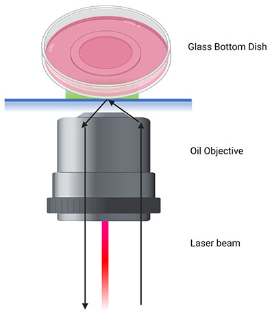
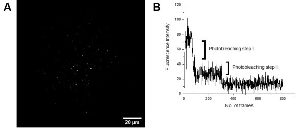
Single molecule imaging and spectroscopy are powerful techniques for the study of a wide range of biological processes including protein assembly and trafficking. However, in vivo single molecule imaging of biomolecules has been challenging because of difficulties associated with sample preparation and technical challenges associated with isolating single proteins within a biological system. Here we provide a detailed protocol to conduct ex vivo single molecule imaging where single transmembrane proteins are isolated by rapidly extracting nanovesicles containing receptors of interest from different regions of the brain and subjecting them to single molecule study by using total internal reflection fluorescence (TIRF) microscopy. This protocol discusses the isolation and separation of brain region specific nanovesicles as well as a detailed method to perform TIRF microscopy with those nanovesicles at the single molecule level. This technique can be applied to study trafficking and stoichiometry of various transmembrane proteins from the central nervous system. This approach can be applied to a wide range of animals that are genetically modified to express a membrane protein-fluorescent protein fusion with a wide range of potential applications in many aspects of neurobiology. Graphic abstract: EX vivo single molecue imaging of membrane receptors.

Keywords: Brain region specific imaging; Membrane proteins; Nanovesicles; Nicotinic acetylcholine receptors; Single molecule imaging; TIRF imaging.


Conflict of interest statement

Competing interestsWe declare no conflict of interest or competitive interest related to this publication.

Figures

Figure 1.
Figure 1.. Set up for Brain Slicing and Region Identification.
A. A mouse brain kept in a 2 mm Zivic brain matrix. B. Blades kept on the brain matrix to make 2 mm thin mouse brain slices. C. An illustration of mouse brain to identify various brain regions from the slices. The anterior part contains the olfactory bulb, and the posterior part is the cerebellum attached to the spinal cord. Slices shown at the bottom contain parts between the olfactory bulb and the cerebellum.
Figure 2.
Figure 2.. Schematic Diagram of GFP containing vesicles immobilized in a dish.
A clean glass bottom dish is coated with biotin-peg-silane, the silane will bind with the glass and biotin will be available to bind with neutravidin. Neutravidin has binding sites to bind with biotin. During washing steps, unbound neutravidin will be washed away while the leftover sites of the bound neutravidin will be used to bind biotinylated anti-GFP antibody. This antibody will bind to the GFP molecule present in the nicotinic acetylcholine receptor of the nanovesicles.
Figure 3.
Figure 3.. Schematic Diagram of TIRF Microscopy on a Glass Bottom Dish.
An oil objective is used where the laser beam is adjusted such that it returns by total internal reflection. The bottom ~200 nm thickness of the dish is illuminated by the evanescent field that originates from total internal reflection on the glass side. This filed allows to image fluorescent single molecule nanovesicles on the bottom surface of the glass bottom dish.
Figure 4.
Figure 4.. Example data from the single molecule imaging of alpha-4 GFP nanovesicles.
A. A TIRF image of spatially isolated nanovesicles containing GFP tethered membrane receptors (scale bar = 5 µm). B. Fluorescence intensity pattern of an individual single molecule from the nanovesicle showing two photobleaching steps (a single molecule region of interest (ROI) is shown in (A)). These photobleaching steps give information about how the alpha-4 beta-2 nAChRs are distributed in the brain.

References

    1. Cano M., Reynaga D. D., Belluzzi J. D., Loughlin S. E. and Leslie F.(2020). Chronic exposure to cigarette smoke extract upregulates nicotinic receptor binding in adult and adolescent rats. Neuropharmacology 181: 108308. - PMC - PubMed
    1. Choi J., Grosely R., Puglisi E. V. and Puglisi J. D.(2019). Expanding single-molecule fluorescence spectroscopy to capture complexity in biology. Curr Opin Struct Biol 58: 233-240. - PMC - PubMed
    1. Fu X., Moonschi F. H., Fox-Loe A. M., Snell A. A., Hopkins D. M., Avelar A. J., Henderson B. J., Pauly J. R. and Richards C. I.(2019). Brain Region Specific Single-Molecule Fluorescence Imaging. Anal Chem 91(15): 10125-10131. - PMC - PubMed
    1. Gage G. J., Kipke D. R. and Shain W.(2012). Whole animal perfusion fixation for rodents. J Vis Exp(65): e3564. - PMC - PubMed
    1. Joo C., Balci H., Ishitsuka Y., Buranachai C. and Ha T.(2008). Advances in single-molecule fluorescence methods for molecular biology. Annu Rev Biochem 77: 51-76. - PubMed

LinkOut - more resources