. 2021 Aug 1;321(2):F207-F224.
doi: 10.1152/ajprenal.00579.2020. Epub 2021 Jun 21.

Differential localization patterns of claudin 10, 16, and 19 in human, mouse, and rat renal tubular epithelia

Affiliations

Differential localization patterns of claudin 10, 16, and 19 in human, mouse, and rat renal tubular epithelia

Caroline Prot-Bertoye et al. Am J Physiol Renal Physiol. .

Abstract

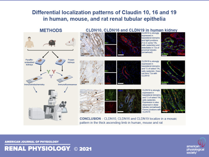
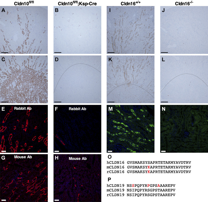
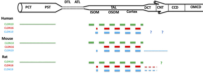
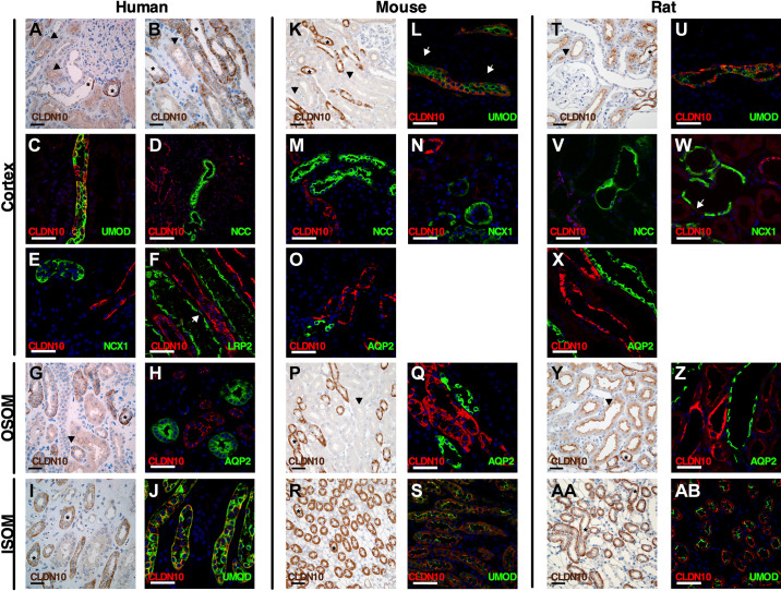
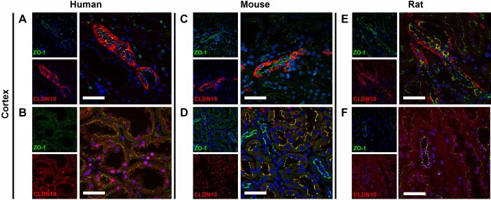
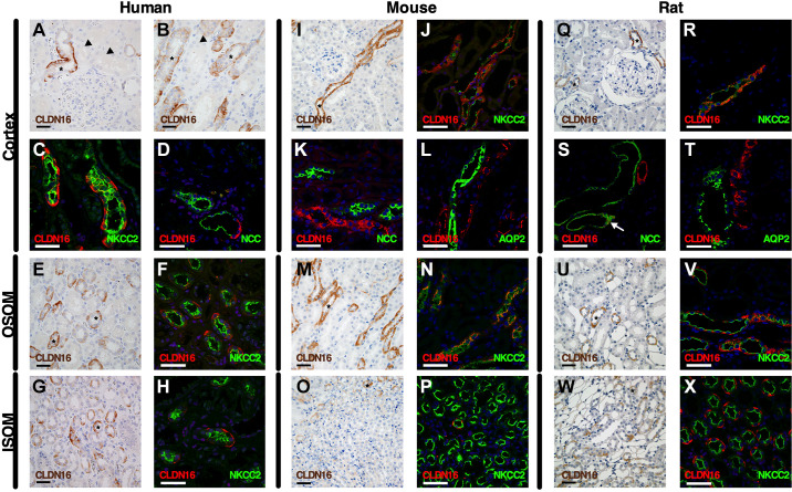
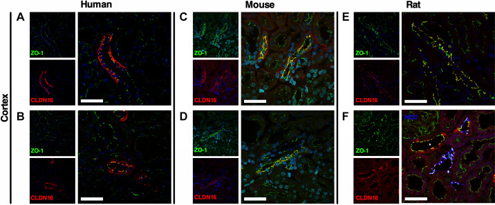
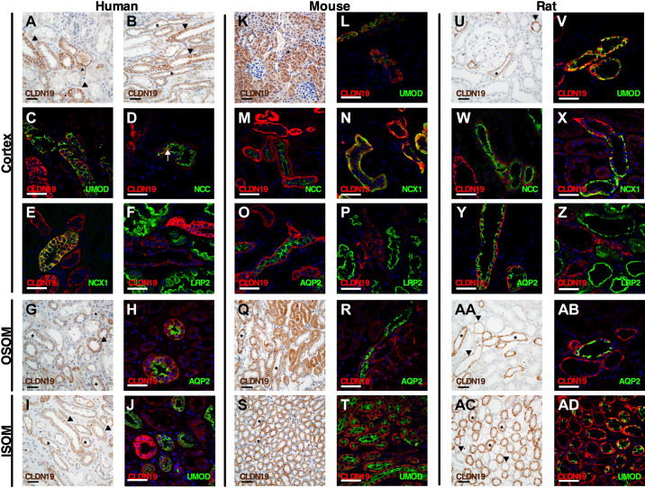
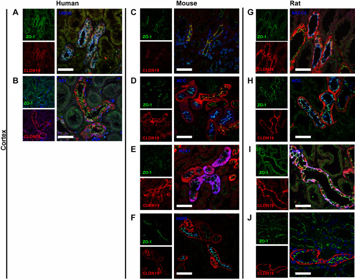
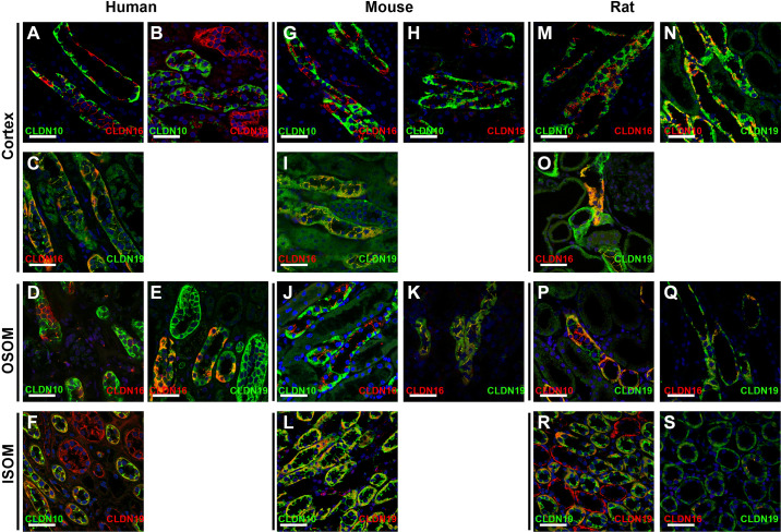
Functional properties of the paracellular pathway depend critically on the set of claudins (CLDN) expressed at the tight junction. Two syndromes are causally linked to loss-of-function mutations of claudins: hypohidrosis, electrolyte imbalance, lacrimal gland dysfunction, ichthyosis, and xerostomia (HELIX) syndrome caused by genetic variations in the CLDN10 gene and familial hypomagnesemia with hypercalciuria and nephrocalcinosis caused by genetic variations in the CLDN16 or CLDN19 genes. All three genes are expressed in the kidney, particularly in the thick ascending limb (TAL). However, localization of these claudins in humans and rodents remains to be delineated in detail. We studied the segmental and subcellular expression of CLDN10, CLDN16, and CLDN19 in both paraffin-embedded and frozen kidney sections from the adult human, mouse, and rat using immunohistochemistry and immunofluorescence, respectively. Here, CLDN10 was present in a subset of medullary and cortical TAL cells, localizing to basolateral domains and tight junctions in human and rodent kidneys. Weak expression was detected at the tight junction of proximal tubular cells. CLDN16 was primarily expressed in a subset of TAL cells in the cortex and outer stripe of outer medulla, restricted to basolateral domains and tight junctional structures in both human and rodent kidneys. CLDN19 predominantly colocalized with CLDN16 in tight junctions and basolateral domains of the TAL but was also found in basolateral and junctional domains in more distal sites. CLDN10 expression at tight junctions almost never overlapped with that of CLND16 and CLDN19, consistent with distinct junctional pathways with different permeation profiles in both human and rodent kidneys.NEW & NOTEWORTHY This study used immunohistochemistry and immunofluorescence to investigate the distribution of claudin 10, 16, and 19 in the human, mouse, and rat kidney. The findings showed distinct junctional pathways in both human and rodent kidneys, supporting the existence of different permeation profiles in all species investigated.

Keywords: epithelium; immunolocalization; kidney; thick ascending limb; tight junction.


Conflict of interest statement

No conflicts of interest, financial or otherwise, are declared by the authors.

Figures

None
Graphical abstract
Figure 1.
Figure 1.
Validation of anticlaudin (CLDN)16 and anti-CLDN10 antibodies (Ab) in wild-type (WT) mice and Cldn16- or Cldn10-specific knockout (KO) mice. A–D: immunohistochemistry for CLDN10 in paraffin-embedded cortical and medullary kidney tissue from WT (Cldn10fl/fl; A and C) and Cldn10 KO mice with kidney-specific deletion (Cldn10fl/fl;Ksp-Cre; B and D). A and B: cortex/outer stripe of the outer medulla (OSOM), C and D: outer medulla. Scale bars = 200 µm. E−H: immunofluorescence for CLDN10 in the outer medulla from WT (E: rabbit anti-CLDN10 antibody and G: mouse anti-CLDN10 antibody) and Cldn10 knockout mice with kidney-specific deletion (F: rabbit anti-CLDN10 antibody and H: mouse anti-CLDN10 antibody). Scale bars = 50 µm. I−L: immunohistochemistry for CLDN16 in paraffin-embedded cortical and medullary kidney tissue from WT (Cldn16+/+) mice [I: cortex/OSOM and K: OSOM/inner stripe of the outer medulla (ISOM)] and Cldn16 KO (Cldn16−/−) mice (J: cortex/OSOM and L: OSOM/ISOM). Scale bars = 200 µm. M and N: immunofluorescence for CLDN16 in frozen cortical kidney tissue from WT (M) and Cldn16 KO mice (N). Scale bars = 50 µm. The line indicates the border between the ISOM and OSOM. O and P: alignments of the parts of the amino acid sequence of CLDN16 (O) and CLDN19 (P) from the human, mouse, and rat used to generate antibodies: anti-CLDN16 antibody was generated against the human epitope and anti-CLDN19 antibody was generated against the mouse epitope. The differences between human and rodent sequences are shown in red.
Figure 2.
Figure 2.
Summary of expression at the tight junction of claudin (CLDN)10, CLDN16, and CLDN19 in human, mouse, and rat renal tubules and collecting ducts. Shown is the relative expression of a given claudin between different segments of the nephron in the same species. The dashed line indicates that some cells do not express the corresponding protein at the tight junction. The dotted line indicates that a minority of cells expresses the corresponding protein at the tight junction. The thickness of the line indicates the relative intensity of expression of the corresponding protein at the tight junction. Relative intensities of expression should not be compared between different claudins expressed by the same species nor between the same claudin expressed by different species. ATL, ascending thin limb; CCD, cortical collecting duct; CNT, connecting tubule; DCT, distal convoluted tubule; DTL, descending thin limb of Henle’s loop; ISOM, inner stripe of the outer medulla; OMCD, outer medullary collecting duct; OSOM, outer stripe of the outer medulla; PCT, proximal convoluted tubule; PST, proximal straight tubule; TAL, thick ascending limb of Henle’s loop. The expression of claudins in the inner medulla was not studied. ?, Expression at the tight junction could not be confirmed nor excluded.
Figure 3.
Figure 3.
Expression of claudin (CLDN)10 in the human, mouse, and rat renal tubule and collecting duct. Immunostaining for CLDN10 in kidney tissue from humans (left), mice (middle), and rats (right) in the cortex, outer stripe of the outer medulla (OSOM), and inner stripe of the outer medulla (ISOM). Paraffin-embedded sections were stained from human (A, B, G, and I), mouse (K, P, and R), and rat (T, Y, and AA) kidneys using anti-CLDN10 antibody. Scale bars = 50 µm. Arrowheads indicate examples of proximal tubular staining. *Thick ascending limb (TAL) of Henle’s loop. Frozen sections were evaluated by colabeling human (C–F, H, and J), mouse (L–O, Q, and S), and rat (U–X, Z, and AB) kidneys using anti-CLDN10 antibody (red) with the following specific segment markers (green) along the renal tubule: megalin/LDL receptor-related protein 2 (LRP2), uromodulin (UMOD), Na+-Cl cotransporter (NCC), Na+-Ca2+ exchanger (NCX1), and aquaporin-2 (AQP2). In F and L, the arrows show weak apical staining in the proximal tubule. This apical proximal tubular staining of CLDN10 is better visualized in Fig. 4, B, D, and E). In W, the arrow indicates weak apical staining in the connecting tubule. Scale bars = 50 µm.
Figure 4.
Figure 4.
Colocalization of claudin (CLDN)10 and zonula occludens-1 (ZO-1) in human, mouse, and rat kidneys. CLDN10 was colocalized with ZO-1 in the human (A and B), mouse (C and D), and rat (E and F) kidney cortex. CLDN10 was expressed at the tight junction in the thick ascending limb of Henle’s loop (A, C, and E) and in proximal tubules (B, D, and F). Scale bars = 50 µm.
Figure 5.
Figure 5.
Expression of claudin (CLDN)16 in the human, mouse, and rat renal tubule and collecting duct. Shown is immunostaining for CLDN16 in kidney tissue from humans (left), mice (middle), and rats (right) in the cortex, outer stripe of the outer medulla (OSOM), and inner stripe of the outer medulla (ISOM). Paraffin-embedded sections were stained in human (A, B, E, and G), mouse (I, M, and O), and rat (Q, U, and W) kidneys using anti-CLDN16 antibody. Scale bars = 50 µm. *Examples of thick ascending limb (TAL) of Henle’s loop. Frozen sections were evaluated by colabeling human (C, D, F, and H), mouse (J –L, N, and P), and rat (R–T, V, and X) kidneys using anti-CLDN16 antibody (red) with the following specific segment markers (green) along the renal tubule: Na+-K+-2Cl cotransporter (NKCC2), Na+-Cl cotransporter (NCC), and aquaporin-2 (AQP2). Since the CLDN16 antibody was raised in mice, we used a TAL marker, the furosemide-sensitive cotransporter (NKCC2), to perform colocalization experiments on frozen sections. In A and B, the arrowheads indicate proximal tubules. In S, the arrow shows CLDN16 expression at the tight junction of the distal convoluted tubule. Scale bars = 50 µm.
Figure 6.
Figure 6.
Colocalization of claudin (CLDN)16 and zonula occludens-1 (ZO-1) in human, mouse, and rat kidneys. CLDN16 and ZO-1 were colocalized in humans (A and B), mice (C and D), and rats (E and F) in the kidney cortex. CLDN16 was expressed at tight junctions in the human, mouse, and rat thick ascending limb of Henle’s loop (A–F) and in the rat distal convoluted tubule (F). Colabeling on kidney sections with the Na+-Cl cotransporter (NCC) was performed. *Thick ascending limb of Henle’s loop. Scale bars = 50 µm.
Figure 7.
Figure 7.
Expression of claudin (CLDN)19 in the human, mouse, and rat renal tubule and collecting duct. Immunostaining for CLDN19 in kidney tissue from humans (left), mice (middle), and rats (right) in the cortex, outer stripe of the outer medulla (OSOM), and inner stripe of the outer medulla (ISOM). Paraffin-embedded sections were stained in human (A, B, G, and I), mouse (K, Q, and S), and rat (U, AA, and AC) kidneys using anti-CLDN19 antibody. Scale bars = 50 µm. *Examples of thick ascending limb of Henle’s loop. Arrowheads show the collecting system. Colabeling on kidney sections from humans (C–F, H, and J), mouse (L–P, R, and T), and rat (V–Z, AB, and AD) kidneys using anti-CLDN19 antibody (red) with the following specific segment markers (green) along the renal tubule was performed: megalin/LDL receptor-related protein 2 (LRP2), uromodulin (UMOD), Na+-Cl cotransporter (NCC), Na+/Ca2+ exchanger (NCX1), and aquaporin-2 (AQP2). In D, the arrow shows the expression of CLDN19 in the distal convoluted tubule. Scale bars = 50 µm.
Figure 8.
Figure 8.
Colocalization of claudin (CLDN)19 and zonula occludens-1 (ZO-1) in human, mouse, and rat kidneys. CLDN19 was colocalized with ZO-1 in human (A and B), mouse (C–F), and rat (G–J) kidneys. Colabeling on kidney sections with uromodulin (UMOD), Na+-K+-2Cl cotransporter (NKCC2), Na+-Cl cotransporter (NCC), Na+/Ca2+ exchanger (NCX1), aquaporin-2 (AQP2), or Cl/HCO3 anion exchanger isoform 1 (AE1) was performed. CLDN19 was expressed at the tight junction (TJ) in the thick ascending limb of Henle’s loop in humans (A), mice (C), and rats (G). CLDN19 was expressed at TJs in the distal convoluted tubule in mice (D) and rats (H). CLDN19 was expressed at a few TJs in the mouse connecting tubule (E) not in the rat connecting tubule (I). CLDN19 was not expressed at TJs in human (B), mouse (F), and rat cortical collecting ducts (J). Scale bars = 50 µm.
Figure 9.
Figure 9.
Colocalization of claudin (CLDN)10, CLDN16, and CLDN19 in the human, mouse, and rat renal tubule and collecting duct. Coimmunostaining for CLDN10 and CLDN16, CLDN10 and CLDN19, and CLDN16 and CLDN19 was performed in kidney tissue from humans (AF, left), mice (GL, middle), and rats (MS, right) in the cortex, outer stripe of the outer medulla (OSOM), and inner stripe of the outer medulla (ISOM). Scale bars = 50 µm.

References

    1. Chen L, Clark JZ, Nelson JW, Kaissling B, Ellison DH, Knepper MA. Renal-tubule epithelial cell nomenclature for single-cell RNA-sequencing studies. J Am Soc Nephrol 30: 1358–1364, 2019. [Erratum in J Am Soc Nephrol 30: 2475, 2019] doi:10.1681/ASN.2019040415. - DOI - PMC - PubMed
    1. Kriz W, Bankir L. A standard nomenclature for structures of the kidney. The Renal Commission of the International Union of Physiological Sciences (IUPS). Kidney Int 33: 1–7, 1988. doi:10.1038/ki.1988.1. - DOI - PubMed
    1. Himmerkus N, Plain A, Marques RD, Sonntag SR, Paliege A, Leipziger J, Bleich M. AVP dynamically increases paracellular Na+ permeability and transcellular NaCl transport in the medullary thick ascending limb of Henle’s loop. Pflugers Arch 469: 149–158, 2017. doi:10.1007/s00424-016-1915-5. - DOI - PubMed
    1. Loupy A, Ramakrishnan SK, Wootla B, Chambrey R, de la Faille R, Bourgeois S, Bruneval P, Mandet C, Christensen EI, Faure H, Cheval L, Laghmani K, Collet C, Eladari D, Dodd RH, Ruat M, Houillier P. PTH-independent regulation of blood calcium concentration by the calcium-sensing receptor. J Clin Invest 122: 3355–3367, 2012. doi:10.1172/JCI57407. - DOI - PMC - PubMed
    1. Morla L, Crambert G, Mordasini D, Favre G, Doucet A, Imbert-Teboul M. Proteinase-activated receptor 2 stimulates Na, K-ATPase and sodium reabsorption in native kidney epithelium. J Biol Chem 283: 28020–28028, 2008. doi:10.1074/jbc.M804399200. - DOI - PMC - PubMed

Publication types

LinkOut - more resources