

Case report of a clinically indolent but morphologically high-grade cutaneous mast cell tumor in an adult: Atypical cutaneous mastocytoma or mast cell sarcoma?
- PMID: 34152029
- PMCID: PMC8638666
- DOI: 10.1111/cup.14088
Case report of a clinically indolent but morphologically high-grade cutaneous mast cell tumor in an adult: Atypical cutaneous mastocytoma or mast cell sarcoma?
Abstract
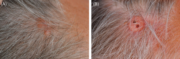
We present a case of an adult male with a solitary mast cell tumor of the skin with unusual nuclear pleomorphism and mitotic activity. The tumor was excised, recurred within 2 years, was reexcised after 4 years and did not recur >6 years after diagnosis. The tumor showed progressive cytonuclear atypia and a high mitotic and proliferation rate by Ki67-staining from the onset. No KIT mutations were identified in the tumor and bone marrow. Serum tryptase levels and a bone marrow aspirate and trephine biopsy were normal. Although the histomorphology of the skin tumor was consistent with mast cell sarcoma, the clinical behavior without systemic progression argued against this diagnosis. The tumor was finally considered as atypical mastocytoma, borderline to mast cell sarcoma. Currently, the patient is in close follow-up and still in complete remission.
Keywords: adult; case reports; mast-cell sarcoma; mastocytoma; mastocytosis.
© 2021 The Authors. Journal of Cutaneous Pathology published by John Wiley & Sons Ltd.
Figures


Similar articles
-
Malignant transformation of mastocytoma developed on skin mastocytosis into cutaneous mast cell sarcoma.Am J Surg Pathol. 2012 May;36(5):779-82. doi: 10.1097/PAS.0b013e31824c0d92. Am J Surg Pathol. 2012. PMID: 22498828
-
Pleomorphic mastocytoma in an adult.J Cutan Pathol. 2018 Feb;45(2):176-179. doi: 10.1111/cup.13080. Epub 2017 Dec 11. J Cutan Pathol. 2018. PMID: 29148588
-
Diagnostic criteria and classification of mastocytosis: a consensus proposal.Leuk Res. 2001 Jul;25(7):603-25. doi: 10.1016/s0145-2126(01)00038-8. Leuk Res. 2001. PMID: 11377686 Review.
-
Mastocytosis: state of the art.Pathobiology. 2007;74(2):121-32. doi: 10.1159/000101711. Pathobiology. 2007. PMID: 17587883 Review.
-
Adult-onset mastocytosis in the skin is highly suggestive of systemic mastocytosis.Mod Pathol. 2014 Jan;27(1):19-29. doi: 10.1038/modpathol.2013.117. Epub 2013 Jun 28. Mod Pathol. 2014. PMID: 23807778
References
-
- Horny H‐P, Akin C, Peterson LC, et al. WHO Classification of Tumours of Haematopoietic and Lymphoid Tissues. 4th ed. Lyon: IARC; 2017. chap 3:61‐69.
-
- Hartmann K, Escribano L, Grattan C, et al. Cutaneous manifestations in patients with mastocytosis: consensus report of the European Competence Network on Mastocytosis; the American Academy of Allergy, Asthma & Immunology; and the European Academy of Allergology and Clinical Immunology. J Allergy Clin Immunol. 2016;137(1):35‐45. - PubMed
-
- Ashinoff R, Soter NA, Freedberg IM. Solitary mastocytoma in an adult. Treatment by excision. J Dermatol Surg Oncol. 1993;19(5):487‐488. - PubMed
-
- Chang IJ, Yang CY, Sung FY, Ng KY. A red‐brown plaque on the nape. Arch Dermatol. 2004;140(10):1275‐1280. - PubMed
Publication types
MeSH terms
Grants and funding
LinkOut - more resources
Full Text Sources
