

Atypical resting state functional connectivity in mild traumatic brain injury
- PMID: 34152089
- PMCID: PMC8413771
- DOI: 10.1002/brb3.2261
Atypical resting state functional connectivity in mild traumatic brain injury
Abstract
Objectives: This study aimed to investigate changes in three intrinsic functional connectivity networks (IFCNs; default mode network [DMN], salience network [SN], and task-positive network [TPN]) in individuals who had sustained a mild traumatic brain injury (mTBI).
Methods: Resting-state functional magnetic resonance imaging (rs-fMRI) data were acquired from 27 mTBI patients with persistent postconcussive symptoms, along with 26 age- and sex-matched controls. These individuals were recruited from a Level-1 trauma center, at least 3 months after a traumatic episode. IFCNs were established based on seed-to-voxel, region-of-interest (ROI) to ROI, and independent component analyses (ICA). Subsequently, we analyzed the relationship between functional connectivity and postconcussive symptoms.
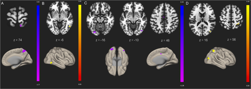
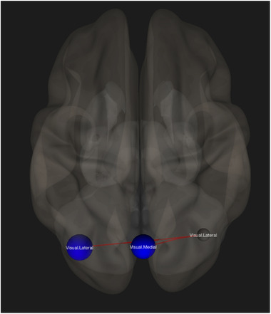
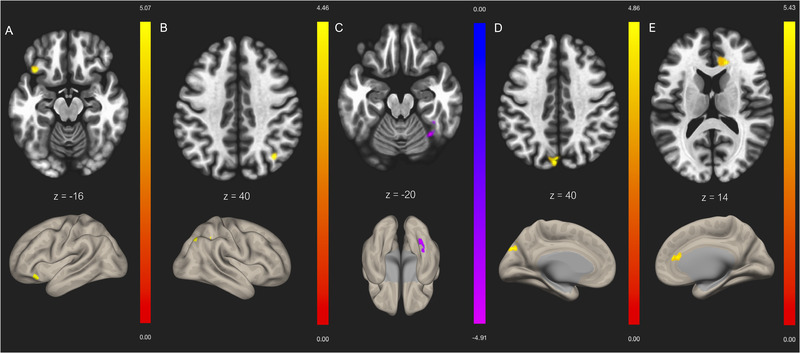
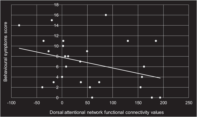
Results: Seed-to-voxel analysis of rs-fMRI demonstrated decreased functional connectivity in the right lateral parietal lobe, part of the DMN, and increased functional connectivity in the supramarginal gyrus, part of the SN. Our TPN showed both hypo- and hyperconnectivity dependent on seed location. Within network hypoconnectivity was observed in the visual network also using group comparison. Using an ICA, we identified altered network functional connectivity in regions within four IFCNs (sensorimotor, visual, DMN, and dorsal attentional). A significant negative correlation between dorsal attentional network connectivity and behavioral symptoms score was also found.
Conclusions: Our findings indicate that rs-fMRI may be of use clinically in order to assess disrupted functional connectivity among IFCNs in mTBI patients. Improved mTBI diagnostic and prognostic information could be especially relevant for athletes looking to safely return to play, as well for individuals from the general population with persistent postconcussive symptoms months after injury, who hope to resume activity.
Keywords: functional magnetic resonance imaging; intrinsic functional connectivity networks; mild traumatic brain injury; postconcussive symptoms; resting state.
© 2021 The Authors. Brain and Behavior published by Wiley Periodicals LLC.
Conflict of interest statement
None of the authors have any conflicts of interest to declare.
Figures
References
-
- Bharath, R. D., Munivenkatappa, A., Gohel, S., Panda, R., Saini, J., Rajeswaran, J., Shukla, D., Bhagavatula, I. D., & Biswal, B. B. (2015). Recovery of resting brain connectivity ensuing mild traumatic brain injury. Frontiers in Human Neuroscience, 9, 513. 10.3389/fnhum.2015.00513 - DOI - PMC - PubMed
-
- Capó‐Aponte, J. E., Jorgensen‐Wagers, K. L., Sosa, J. A., Walsh, D. V., Goodrich, G. L., Temme, L. A., & Riggs, D. W. (2017). Visual dysfunctions at different stages after blast and non‐blast mild traumatic. Brain Injury: Optometry and Vision Science, 94(1), 7–15. 10.1097/OPX.0000000000000825 - DOI - PubMed
-
- Chen, J.‐K., Johnston, K. M., Collie, A., McCrory, P., & Ptito, A. (2007). A validation of the post concussion symptom scale in the assessment of complex concussion using cognitive testing and functional MRI. Journal of Neurology, Neurosurgery & Psychiatry, 78(11), 1231–1238. 10.1136/jnnp.2006.110395 - DOI - PMC - PubMed
-
- Chen, J.‐K., Johnston, K. M., Petrides, M., & Ptito, A. (2008). Recovery from mild head injury in sports: Evidence from serial functional magnetic resonance imaging studies in male athletes. Clinical Journal of Sport Medicine, 18(3), 241. - PubMed
Publication types
MeSH terms
LinkOut - more resources
Full Text Sources
Medical
Research Materials
Miscellaneous
