Review
. 2021 Jun 20:90:507-534.
doi: 10.1146/annurev-biochem-081720-023244.

The Form and Function of PIEZO2

Affiliations
Review

The Form and Function of PIEZO2

Marcin Szczot et al. Annu Rev Biochem. .

Abstract

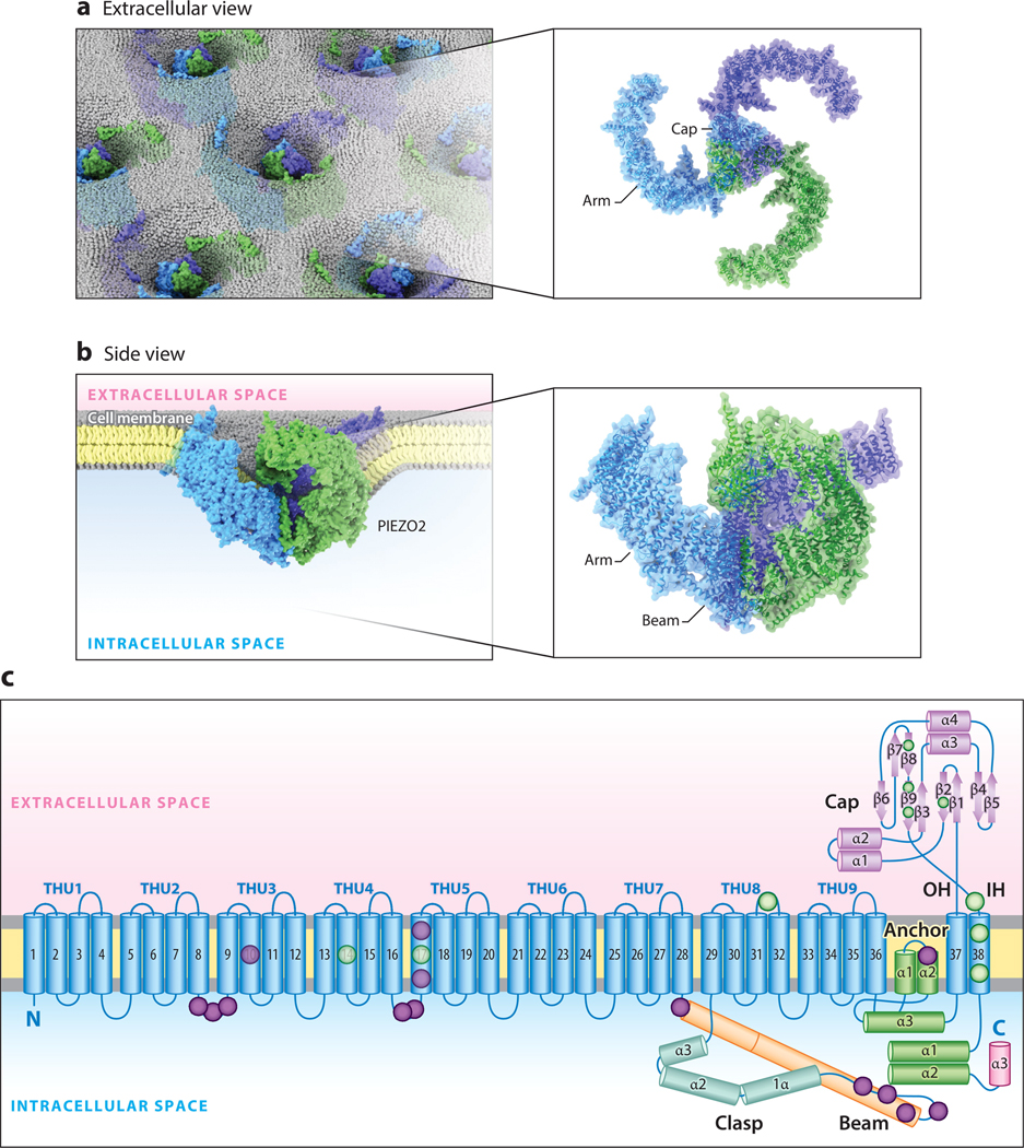
Mechanosensation is the ability to detect dynamic mechanical stimuli (e.g., pressure, stretch, and shear stress) and is essential for a wide variety of processes, including our sense of touch on the skin. How touch is detected and transduced at the molecular level has proved to be one of the great mysteries of sensory biology. A major breakthrough occurred in 2010 with the discovery of a family of mechanically gated ion channels that were coined PIEZOs. The last 10 years of investigation have provided a wealth of information about the functional roles and mechanisms of these molecules. Here we focus on PIEZO2, one of the two PIEZO proteins found in humans and other mammals. We review how work at the molecular, cellular, and systems levels over the past decade has transformed our understanding of touch and led to unexpected insights into other types of mechanosensation beyond the skin.

Keywords: PIEZO2; ion channel; mechanosensation; mechanotransduction; proprioception; somatosensation; touch.


Figures

Figure 1
Figure 1
The structure of mouse PIEZO2. (a) A top-down illustration of mouse PIEZO2 channels in the membrane as viewed from outside the cell. (b) A side-view illustration of a PIEZO2 channel curving the plasma membrane. (c) Ribbon diagram of one blade of PIEZO2 highlighting key functional domains. Lavender and green circles indicate approximate reported locations of human loss-of-function and gain-of-function variants, respectively. Structure adapted with permission from Reference 48. Abbreviations: α, α-helix; β, β-pleated sheet; IH, inner helix of the ion-conducting pore; OH, outer helix of the ion-conducting pore; THU, transmembrane helical unit.
Figure 2
Figure 2
The expression of PIEZO2 in mouse sensory neuron classes. Single-cell RNA sequencing data from four distinct peripheral ganglia are shown: dorsal root, trigeminal, jugular, and nodose. For each ganglion, transcriptomic cell classes are given in the UMAP plots with PIEZO2 expression level visualized and color coded by high expression (green) or no expression (black). (a) Single-cell sequencing from isolated dorsal root ganglia neurons across development, with the earliest embryonic time points at the center of the UMAP and adult neurons at the outer edges (100). (b) Single-nucleus sequencing of trigeminal ganglia. Note that the more prominent representation of Aβ-LTMRs is likely to be the result of the nuclear isolation method (106), which better captures large-diameter neuron subtypes that are normally lost during single-cell isolation. (c) Single-cell sequencing of the vagal complex shows that cell classes in the jugular ganglia are transcriptomically analogous to those found in the dorsal root and trigeminal ganglia. (d) The nodose ganglion, on the other hand, is comprised of transcriptomically and functionally unique sensory neurons (105). The labels for classes of neurons with high PIEZO2 expression are boxed, while those with low or no expression are in italics. Panel a adapted with permission from Reference 100, panels b and d adapted with permission from Reference 105, and panel c adapted from Reference 106. Abbreviations: LTMR, low-threshold mechanoreceptor; UMAP, uniform manifold approximation and projection.
Figure 3
Figure 3
Biological systems in which PIEZO2 transduction is involved. The left column summarizes the clinical phenotypes reported in PIEZO2 deficiency syndrome. The right column describes the physiological roles of PIEZO2 in mouse models that may underlie each phenotype. Abbreviations: DRG, dorsal root ganglion; LTMR, low-threshold mechanoreceptor; SA1-LTMR, slowly adapting type I Aβ-LTMR.
Figure 4
Figure 4
Adapting single-cell mechanical stimulation methods to high-throughput formats. (Left) Existing methods for applying mechanical force to the cell membrane. For reference, a graphic depiction of a single PIEZO2 channel is shown in dark orange spanning the cell membrane. (Right) Potential strategies for adapting each method for use in high-throughput screening platforms. Yellow arrows indicate the direction of mechanical force in each assay.

References

    1. Iggo A, Andres KH. 1982. Morphology of cutaneous receptors. Annu. Rev. Neurosci. 5:1–31 - PubMed
    1. Sherrington C 1906. The Integrative Action of the Nervous System. New Haven: Yale Univ. Press
    1. Perl E 1968. Myelinated afferent fibres innervating the primate skin and their response to noxious stimuli. J. Physiol. 197:593–615 - PMC - PubMed
    1. Burgess P, Petit D, Warren RM. 1968. Receptor types in cat hairy skin supplied by myelinated fibers. J. Neurophysiol. 31:833–48 - PubMed
    1. Brown A, Iggo A. 1967. A quantitative study of cutaneous receptors and afferent fibres in the cat and rabbit. J. Physiol. 193:707–33 - PMC - PubMed

Publication types

LinkOut - more resources