Review
. 2021 Jun 20:90:431-450.
doi: 10.1146/annurev-biochem-081720-020121.

An Overview of Microcrystal Electron Diffraction (MicroED)

Affiliations
Review

An Overview of Microcrystal Electron Diffraction (MicroED)

Xuelang Mu et al. Annu Rev Biochem. .

Abstract

The bedrock of drug discovery and a key tool for understanding cellular function and drug mechanisms of action is the structure determination of chemical compounds, peptides, and proteins. The development of new structure characterization tools, particularly those that fill critical gaps in existing methods, presents important steps forward for structural biology and drug discovery. The emergence of microcrystal electron diffraction (MicroED) expands the application of cryo-electron microscopy to include samples ranging from small molecules and membrane proteins to even large protein complexes using crystals that are one-billionth the size of those required for X-ray crystallography. This review outlines the conception, achievements, and exciting future trajectories for MicroED, an important addition to the existing biophysical toolkit.

Keywords: MicroED; cryo-EM; cryo–electron microscopy; crystallography; microcrystal electron diffraction; proteins; structures.


Figures

Figure 1
Figure 1
The four modalities of cryo–electron microscopy (cryo-EM). (a) A model of the kinetochore assembly by tomography. Panel adapted from Reference with permission from the American Association for the Advancement of Science (AAAS). (b) Structure of a 17-kDa protein, DARPin, on a scaffold by single-particle analysis. Panel adapted from Reference with permission from AAAS. (c) The structure of bacteriorhodopsin determined by electron crystallography of two-dimensional (2D) crystals. Panel adapted with permission from Reference . (d) The structure of catalase determined by microcrystal electron diffraction (MicroED) of 3D crystals. Panel adapted with permission from Reference .
Figure 2
Figure 2
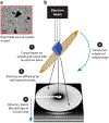
An overview of microcrystal electron diffraction (MicroED). (a) An image of an electron microscope grid in bright field during screening for microcrystals. (b) Once located, a microcrystal is exposed to an electron beam in diffraction mode while being continuously rotated on the sample stage. The diffraction data are then collected as a movie using a fast camera.
Figure 3
Figure 3
Examples of structures of small-molecule chemical compounds solved by microcrystal electron diffraction (MicroED). Structures of carbamazepine, MBBF4, Grippostad, and ITIC-Th are shown.
Figure 4
Figure 4
Focused ion beam (FIB) milling of crystals for microcrystal electron diffraction (MicroED). (a) Image of select proteinase K crystals at high magnification before milling. The arrow indicates the crystal that was milled in panel c. (b) FIB image of select crystal from panel a after milling the top of the crystal. (c) FIB image of crystal after milling and cleaning both the top and bottom of the crystal, leaving a lamella indicated by an arrow. Figure reproduced with permission from Reference .
Figure 5
Figure 5
Examples of protein structures solved by MicroED that were challenging by other techniques. (a) Structure of α-synuclein NACore (66) (PDB ID: 4ZNN). (b) MicroED structures of the membrane proteins rendered as cyan and green ribbons for Ca2+ ATPase (PDB ID: 3J7T/U) (64) and NaK (PDB ID: 6CPV) (63), respectively. (c) MicroED structure of the TGF-βm:TβRII protein-protein complex (PDB ID: 5TY4) (72). Protein rendered as pink and magenta ribbons for TGF-βm and TβRII, respectively. Abbreviations: MicroED, microcrystal electron diffraction; PDB ID, Protein Data Bank identifier.
Figure 6
Figure 6
The structure of the HIV GAG–bevirimat complex solved by microcrystal electron diffraction (MicroED) (104). (a) Overall architecture of HIV GAG-bevirimat complex. (b) Expanded view of the bevirimat binding site in the pore of HIV GAG. Both panels reproduced with permission from Reference .
Figure 7
Figure 7
Brucine, 1-methyl-2-indanone, thiostrepton, and 3-thiaGlu are examples of natural products with structures determined by microcrystal electron diffraction (MicroED).
Figure 8
Figure 8
Radiation damage in microcrystal electron diffraction (MicroED) (70). (a) Proteinase K recorded at various exposure rates. (b) Volume and B-factor were averaged across all the crystals at each exposure for proteinase K. (c) Density loss in arbitrary units for all the amino acids, ligands, and ions present in the refined models of proteinase K. All panels adapted with permission from Reference .
Figure 9
Figure 9
Expanding the use of direct electron detectors and mitigating radiation damage in microcrystal electron diffraction (MicroED) (124). (a) The exposure dependency of the completeness of proteinase K. The dotted horizontal line marks 95% completeness. (b) The density around the two disulfide bonds for the considered cameras for proteinase K. Both panels adapted with permission from Reference .
Figure 10
Figure 10
Identification of heavy metal site and model building by radiation-induced damage for phasing. (a) Fourier difference maps between the damaged and undamaged structure of GSNQNNF using the phases of 6CLI at 2.5 Å resolution contoured at 3σ level. (b) The map using the initial phases extended to 1.4 Å resolution. (c) Density maps at 1.0σ contour for intermediate building steps. (d) The final structure. All panels adapted with permission from Reference .

References

    1. Dubochet J, Booyl FP, Freeman R, Jones AV, Walter CA. 1981. Low temperature electron microscopy. Ann. Rev. Biophys 10:133–49 - PubMed
    1. Taylor KA, Glaeser RM. 1974. Electron diffraction of frozen, hydrated protein crystals. Science 186:1036–37 - PubMed
    1. Wang L, Sigworth FJ. 2006. Cryo-EM and single particles. Physiology 21:13–18 - PubMed
    1. Bai XC, Fernandez IS, McMullan G, Scheres SHW. 2013. Ribosome structures to near-atomic resolution from thirty thousand cryo-EM particles. eLife 2:e00461. - PMC - PubMed
    1. Herzik MA, Wu M, Lander GC. 2017. Achieving better-than-3-Å resolution by single-particle cryo-EM at 200 keV. Nat. Methods 14:1075–78 - PMC - PubMed

Publication types

MeSH terms