. 2021 Jun 29;12(3):e0130221.
doi: 10.1128/mBio.01302-21. Epub 2021 Jun 22.

The Carbapenemase BKC-1 from Klebsiella pneumoniae Is Adapted for Translocation by Both the Tat and Sec Translocons

Affiliations

The Carbapenemase BKC-1 from Klebsiella pneumoniae Is Adapted for Translocation by Both the Tat and Sec Translocons

Manasa Bharathwaj et al. mBio. .

Abstract

The cell envelope of Gram-negative bacteria consists of two membranes surrounding the periplasm and peptidoglycan layer. β-Lactam antibiotics target the periplasmic penicillin-binding proteins that synthesize peptidoglycan, resulting in cell death. The primary means by which bacterial species resist the effects of β-lactam drugs is to populate the periplasmic space with β-lactamases. Resistance to β-lactam drugs is spread by lateral transfer of genes encoding β-lactamases from one species of bacteria to another. However, the resistance phenotype depends in turn on these "alien" protein sequences being recognized and exported across the cytoplasmic membrane by either the Sec or Tat protein translocation machinery of the new bacterial host. Here, we examine BKC-1, a carbapenemase from an unknown bacterial source that has been identified in a single clinical isolate of Klebsiella pneumoniae. BKC-1 was shown to be located in the periplasm, and functional in both K. pneumoniae and Escherichia coli. Sequence analysis revealed the presence of an unusual signal peptide with a twin arginine motif and a duplicated hydrophobic region. Biochemical assays showed this signal peptide directs BKC-1 for translocation by both Sec and Tat translocons. This is one of the few descriptions of a periplasmic protein that is functionally translocated by both export pathways in the same organism, and we suggest it represents a snapshot of evolution for a β-lactamase adapting to functionality in a new host. IMPORTANCE Bacteria can readily acquire plasmids via lateral gene transfer (LGT). These plasmids can carry genes for virulence and antimicrobial resistance (AMR). Of growing concern are LGT events that spread β-lactamases, particularly carbapenemases, and it is important to understand what limits this spread. This study provides insight into the sequence features of BKC-1 that exemplify the limitations on the successful biogenesis of β-lactamases, which is one factor limiting the spread of AMR phenotypes by LGT. With a very simple evolutionary adaptation, BKC-1 could become a more effective carbapenemase, underscoring the need to understand the evolution, adaptability, and functional assessment of newly reported β-lactamases rapidly and thoroughly.

Keywords: Tat pathway; antimicrobial resistance; beta-lactamases; evolution; periplasm; protein secretion; signal peptide; β-lactamase.


Figures
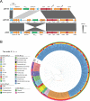
FIG 1
FIG 1
Comparison of the genetic environment of BKC-1. (A) The blaBKC-1 gene is carried on an RSF1010 plasmid, p60136 (59), the sequence of which was aligned with two related IncQ plasmids from K. pneumoniae (pKPC05 with 87.58% overall sequence identity and pB29 with 84.61% overall sequence identity) using Easyfig 2.2.5. Arrows represent genes as follows: β-lactamase encoding genes (green); replication-related genes (red); other genes of known function (orange); and genes of unknown function (blue). (B) A global phylogeny of class A β-lactamases was constructed by sequence comparison of class A β-lactamases from BLDB. The sequences displayed were retrieved from BLDB allowing for at least 40% sequence identity. The position of BKC-1 is denoted in bold. GPC-1, a β-lactamase identified in P. aeruginosa, showed the greatest amino acid sequence identity to BKC-1 (77%), while the next closest relationship (63% sequence identity) was with the chromosomally encoded β-lactamase (PAD-1) from P. desertii. The four rings that designate features in the phylogeny are as indicated in the legend.
FIG 2
FIG 2
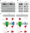
A unique signal sequence targets BKC-1 into the periplasm. (A) The position and sequence of the signal peptide encoded in blaBKC-1. The twin arginine motif (underlined), 32-residue h-region (red), and cleavage site (AGA-AT) are indicated. (B) Protein sequence alignment of BKC-1 from K. pneumoniae, GPC-1 from P. aeruginosa, and PAD-1 from P. desertii. Red indicates identical residues; yellow indicates conserved residues. (C and D) Cell lysates from E. coli transformed with pJPCmR (–) or pJPBKC-1His encoding C-terminally His6-tagged BKC-1 (+) were fractionated (C) or TSE-extracted (D) and analyzed by SDS-PAGE and immunoblotting using antibodies raised against BKC-1, BamA (membrane protein control), or SurA (periplasmic protein control). Asterisks indicate the slower-migrating nonspecific protein present in SurA immunoblots.
FIG 3
FIG 3
The unique signal sequence of BKC-1 is required for efficient Tat-independent translocation into the periplasm. (A and B) Whole-cell lysates were prepared from wild-type E. coli BW25113 or its isogenic ΔtatC mutant to compare levels of BKC-1 with other β-lactamases (A) or its BKC-1A derivative that has a shorter signal peptide (B). pJPCmR or its derivative vectors containing C-terminally His6-tagged β-lactamases proteins were used to synthesize the indicated protein of interest (above immunoblot). The extracts were analyzed by SDS-PAGE and immunoblotting using antibodies raised against the indicated proteins (right of immunoblot). SurA and F1β serve as loading controls. Asterisks indicate precursor protein forms that migrate slower by SDS-PAGE. (C) The proposed model for the function and translocation of BKC-1 across the inner membrane (IM) in the presence and absence of TatC. The topological compartments of the periplasm and outer membrane (OM) are indicated. The unfolded and folded forms of BKC-1 (red) are shown and the rectangle with twin arginines (RR) represents its signal peptide. In the periplasm, both translocated forms of BKC-1 work together to hydrolyze β-lactam antibiotics, but in the absence of TatC, there is a decreased number of BKC-1 that can enter the periplasm.

References

    1. Tuller T, Girshovich Y, Sella Y, Kreimer A, Freilich S, Kupiec M, Gophna U, Ruppin E. 2011. Association between translation efficiency and horizontal gene transfer within microbial communities. Nucleic Acids Res 39:4743–4755. doi:10.1093/nar/gkr054. - DOI - PMC - PubMed
    1. Dalbey RE, Kuhn A. 2012. Protein traffic in Gram-negative bacteria—how exported and secreted proteins find their way. FEMS Microbiol Rev 36:1023–1045. doi:10.1111/j.1574-6976.2012.00327.x. - DOI - PubMed
    1. Bogumil D, Dagan T. 2012. Cumulative impact of chaperone-mediated folding on genome evolution. Biochemistry 51:9941–9953. doi:10.1021/bi3013643. - DOI - PubMed
    1. Ochman H, Lawrence JG, Groisman EA. 2000. Lateral gene transfer and the nature of bacterial innovation. Nature 405:299–304. doi:10.1038/35012500. - DOI - PubMed
    1. Ruzzini AC, Clardy J. 2016. Gene flow and molecular innovation in bacteria. Curr Biol 26:R859–R864. doi:10.1016/j.cub.2016.08.004. - DOI - PMC - PubMed

Publication types

MeSH terms

LinkOut - more resources