. 2021 Jun 29;118(26):e2102787118.
doi: 10.1073/pnas.2102787118.

C1q binding to surface-bound IgG is stabilized by C1r2s2 proteases

Affiliations

C1q binding to surface-bound IgG is stabilized by C1r2s2 proteases

Seline A Zwarthoff et al. Proc Natl Acad Sci U S A. .

Abstract

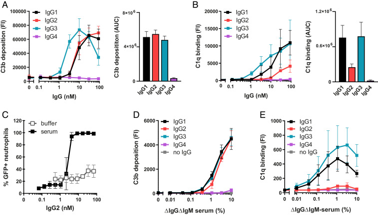
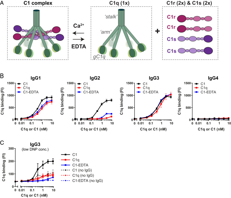
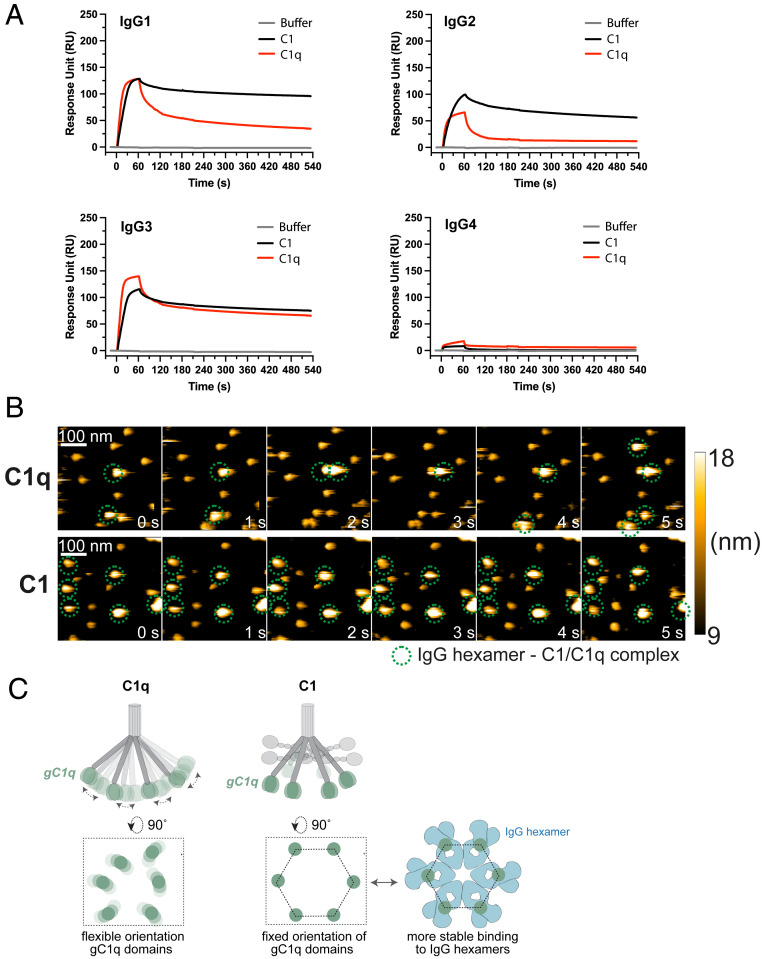
Complement is an important effector mechanism for antibody-mediated clearance of infections and tumor cells. Upon binding to target cells, the antibody's constant (Fc) domain recruits complement component C1 to initiate a proteolytic cascade that generates lytic pores and stimulates phagocytosis. The C1 complex (C1qr2s2) consists of the large recognition protein C1q and a heterotetramer of proteases C1r and C1s (C1r2s2). While interactions between C1 and IgG-Fc are believed to be mediated by the globular heads of C1q, we here find that C1r2s2 proteases affect the capacity of C1q to form an avid complex with surface-bound IgG molecules (on various 2,4-dinitrophenol [DNP]-coated surfaces and pathogenic Staphylococcus aureus). The extent to which C1r2s2 contributes to C1q-IgG stability strongly differs between human IgG subclasses. Using antibody engineering of monoclonal IgG, we reveal that hexamer-enhancing mutations improve C1q-IgG stability, both in the absence and presence of C1r2s2 In addition, hexamer-enhanced IgGs targeting S. aureus mediate improved complement-dependent phagocytosis by human neutrophils. Altogether, these molecular insights into complement binding to surface-bound IgGs could be important for optimal design of antibody therapies.

Keywords: C1; IgG hexamerization; IgG subclasses; Staphylococcus aureus; complement.


Conflict of interest statement

Competing interest statement: A.K., J.A.G.v.S., P.W.H.I.P., K.P.M.v.K., F.J.B., J. Schuurman, and S.H.M.R. are coinventors on a patent describing antibody therapies against S. aureus.

Figures

Fig. 1.
Fig. 1.
IgG-mediated complement activation does not always correlate with detection of C1q. (A and B) C3b deposition (A) and C1q binding (B) on anti-WTA IgG-labeled S. aureus Wood46 bacteria upon incubation with 5% or, respectively, 1% ΔIgGΔIgM serum as determined by flow cytometry. The data represent mean FI ± SD (Left) or mean area under the curve (AUC) ± SD (Right) of three independent experiments. AUC of C3b and C1q binding curves was determined after subtraction of a 2,500 (C3b) or 200 (C1q) baseline, respectively. (C) Phagocytosis of fluorescently labeled S. aureus Wood46 in either RPMI buffer or 1% ΔIgGΔIgM serum supplemented with anti-WTA IgG2 and human neutrophils. Bacterial uptake was quantified by flow cytometry and displayed as the percentage of GFP-positive neutrophils. The data represent mean ± SD of three independent experiments. (D and E) C3b deposition (D) and detection of C1q (E) on beads coated with 1 µg/mL DNP upon incubation with ΔIgGΔIgM serum and human monoclonal anti-DNP IgG (20 nM). Deposition of C3b and C1q molecules on the beads was determined by flow cytometry. The data represent geometric mean ± SD of three independent experiments.
Fig. 2.
Fig. 2.
C1r2s2 proteases enhance the binding of C1q to target-bound IgG. (A) Schematic presentation of C1 consisting of the recognition molecule C1q in complex with a tetramer of proteases C1r and C1s (C1r2s2). C1q consists of six polypeptide chains that come together in an N-terminal stalk. At their C terminus, all six chains end in a globular domain (gC1q) that recognizes Fc domains of IgM and clustered IgGs. The C1r2s2 tetramer associates to the collagen arms of C1q via Ca2+-dependent interactions (56). The proteases dissociate from C1q in the presence of EDTA, a calcium chelator. (B) Binding of different forms of purified C1 to DNP beads (1 µg/mL DNP) coated with 20 nM anti-DNP IgG1-4. “C1” indicates the fully assembled C1 complex (= C1qr2s2), “C1q” is the recognition molecule C1q only, “C1-EDTA” sample consists of C1q, C1r, and C1s, but the proteases are not attached to C1q (because 10 mM EDTA disrupts the Ca2+-dependent association between proteases and C1q). (C) Binding of different forms of purified C1 to IgG3-labeled beads coated with 0.003 µg/mL (∼300-fold lower than in B). The dotted lines show aspecific binding of the C1q molecules in absence of IgG3. (B and C) Bound C1q was detected by polyclonal anti-C1q antibodies and flow cytometry. The data represent mean ± SD of three independent experiments.
Fig. 3.
Fig. 3.
C1r2s2 proteases enhance the stability of surface-bound C1q–IgG complexes. (A) SPR experiment showing binding of purified C1 or C1q to sensor chips with immobilized DNP coated with 20 nM anti-DNP IgG. C1 or C1q was injected for 60 s to allow association, after which the injection was stopped and dissociation was monitored. Representative of two independent experiments. SPR responses were normalized to account for the molecular weight difference between C1 (766 kDa) and C1q (410 kDa). RU, response units. (B) HS-AFM image sequence of anti-DNP IgG1-RGY in complex with C1q in the presence of C1q in solution (Upper; taken from Movie S1) and C1 in the absence of C1 in solution (Lower; taken from Movie S2). The depicted height scale is relative to the membrane surface. The heights of the respective complexes were 12.2 ± 0.6 (anti-DNP IgG1-RGY) (17), 20.2 ± 1.5 (anti-DNP IgG1-RGY + C1q), and 18.5 ± 1.7 nm (anti-DNP IgG1-RGY + C1). (C) gC1q domains mediate binding to IgG. In C1q, the gC1q domains are flexible. We hypothesize that in C1 the associated C1r2s2 proteases fix the collagen arms and thereby orient the gC1q domains in a hexagon-like platform that favors binding to (hexameric) IgG clusters.
Fig. 4.
Fig. 4.
Removal of C1r2s2 proteases from surface-bound C1–IgG complexes can result in C1q dissociation. (A) Detection of C1q on IgG-coated DNP beads that were first labeled with purified C1 and subsequently incubated with 10 mM EDTA or 200 nM C1-INH to remove C1r and C1s proteases. The data represent geometric mean ± SD of three independent experiments. Unpaired Student’s t test (buffer versus EDTA; buffer versus C1-INH); *P < 0.05, all other conditions not significant. (B) Schematic cartoon of our hypothesis that C1r2s2 dissociation by C1-INH can result in C1q dislodgement depending on the stability of the C1q–IgG complex. Our data suggest that removal of the C1r and C1s proteases from surface-bound C1 by C1-INH can result in two situations: 1) C1q dissociates from the surface-bound IgGs in the case the remaining C1q–IgG complexes are unstable (e.g., for C1q–IgG2 complexes) or 2) C1q remains bound since it has formed a stable interaction with the surface-bound IgGs.
Fig. 5.
Fig. 5.
Enhanced IgG oligomerization stabilizes C1q–IgG interactions. (A and B) Binding of purified C1q (A) or C1 (B) to DNP beads (1 µg/mL DNP) labeled with 20 nM anti-DNP IgG, either wild-type or containing hexamer-enhancing mutations E430G or E345K. (C) Detection of C1q on DNP-beads (1 µg/mL DNP) after incubation with 1% ΔIgGΔIgM serum supplemented with wild-type or mutated (E430G) anti-DNP IgG. (AC) C1q was detected by flow cytometry. The data represent geometric mean ± SD of three independent experiments.
Fig. 6.
Fig. 6.
Introduction of hexamer-enhancing mutation E430G in anti-WTA can enhance complement-dependent phagocytosis of S. aureus. (A) C3b deposition on S. aureus Wood46 after incubation of bacteria in 5% ΔIgGΔIgM serum supplemented with anti-WTA IgG (wild-type or E430G mutant). The data represent mean ± SD of three independent experiments. (B) Phagocytosis in the absence and presence of complement. Phagocytosis of fluorescently labeled S. aureus Wood46 in either RPMI buffer or 1% ΔIgGΔIgM serum supplemented with anti-WTA IgG (wild-type or E430G mutant) and human neutrophils. Bacterial uptake was quantified by flow cytometry and displayed as the number of GFP-positive neutrophils relative to IgG1 wild type. The data represent relative mean ± SD of three independent experiments. Phagocytosis data shown for wild-type IgG2 (buffer and serum condition) are identical to the data shown in Fig. 1C.

References

    1. Lu L. L., Suscovich T. J., Fortune S. M., Alter G., Beyond binding: Antibody effector functions in infectious diseases. Nat. Rev. Immunol. 18, 46–61 (2018). - PMC - PubMed
    1. Walport M. J., Complement. First of two parts. N. Engl. J. Med. 344, 1058–1066 (2001). - PubMed
    1. Gros P., Milder F. J., Janssen B. J. C., Complement driven by conformational changes. Nat. Rev. Immunol. 8, 48–58 (2008). - PubMed
    1. Papanastasiou M., et al., Structural implications for the formation and function of the complement effector protein iC3b. J. Immunol. 198, 3326–3335 (2017). - PMC - PubMed
    1. Ricklin D., Hajishengallis G., Yang K., Lambris J. D., Complement: A key system for immune surveillance and homeostasis. Nat. Immunol. 11, 785–797 (2010). - PMC - PubMed

Publication types

LinkOut - more resources