

Sequence signatures of two public antibody clonotypes that bind SARS-CoV-2 receptor binding domain
- PMID: 34155209
- PMCID: PMC8217500
- DOI: 10.1038/s41467-021-24123-7
Sequence signatures of two public antibody clonotypes that bind SARS-CoV-2 receptor binding domain
Abstract
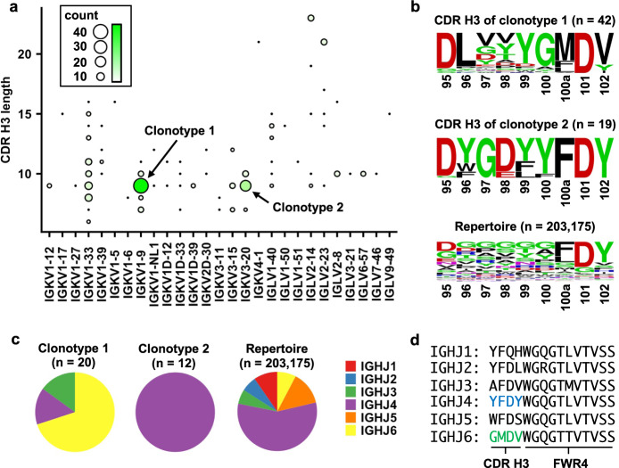
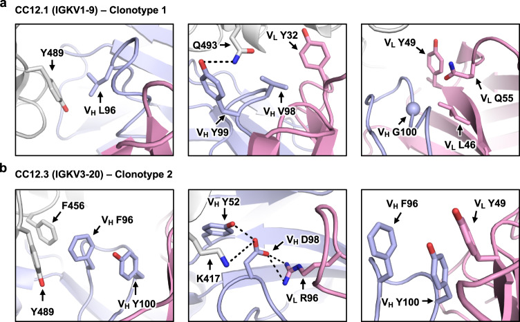
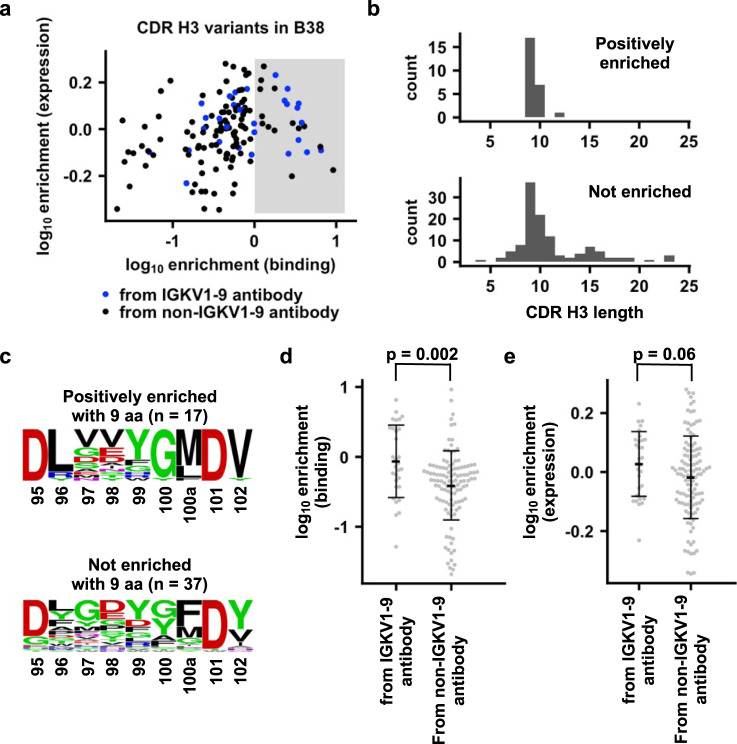
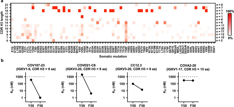
Since the COVID-19 pandemic onset, the antibody response to SARS-CoV-2 has been extensively characterized. Antibodies to the receptor binding domain (RBD) on the spike protein are frequently encoded by IGHV3-53/3-66 with a short complementarity-determining region (CDR) H3. Germline-encoded sequence motifs in heavy chain CDRs H1 and H2 have a major function, but whether any common motifs are present in CDR H3, which is often critical for binding specificity, is not clear. Here, we identify two public clonotypes of IGHV3-53/3-66 RBD antibodies with a 9-residue CDR H3 that pair with different light chains. Distinct sequence motifs on CDR H3 are present in the two public clonotypes that seem to be related to differential light chain pairing. Additionally, we show that Y58F is a common somatic hypermutation that results in increased binding affinity of IGHV3-53/3-66 RBD antibodies with a short CDR H3. These results advance understanding of the antibody response to SARS-CoV-2.
Conflict of interest statement
The authors declare no competing interests.
Figures
Update of
-
Sequence signatures of two IGHV3-53/3-66 public clonotypes to SARS-CoV-2 receptor binding domain.bioRxiv [Preprint]. 2021 Jan 27:2021.01.26.428356. doi: 10.1101/2021.01.26.428356. bioRxiv. 2021. Update in: Nat Commun. 2021 Jun 21;12(1):3815. doi: 10.1038/s41467-021-24123-7. PMID: 33532781 Free PMC article. Updated. Preprint.
Similar articles
-
Sequence signatures of two IGHV3-53/3-66 public clonotypes to SARS-CoV-2 receptor binding domain.bioRxiv [Preprint]. 2021 Jan 27:2021.01.26.428356. doi: 10.1101/2021.01.26.428356. bioRxiv. 2021. Update in: Nat Commun. 2021 Jun 21;12(1):3815. doi: 10.1038/s41467-021-24123-7. PMID: 33532781 Free PMC article. Updated. Preprint.
-
An Alternative Binding Mode of IGHV3-53 Antibodies to the SARS-CoV-2 Receptor Binding Domain.Cell Rep. 2020 Oct 20;33(3):108274. doi: 10.1016/j.celrep.2020.108274. Epub 2020 Sep 29. Cell Rep. 2020. PMID: 33027617 Free PMC article.
-
Paired heavy- and light-chain signatures contribute to potent SARS-CoV-2 neutralization in public antibody responses.Cell Rep. 2021 Oct 5;37(1):109771. doi: 10.1016/j.celrep.2021.109771. Epub 2021 Sep 28. Cell Rep. 2021. PMID: 34587480 Free PMC article.
-
Mutations in the SARS-CoV-2 spike receptor binding domain and their delicate balance between ACE2 affinity and antibody evasion.Protein Cell. 2024 May 28;15(6):403-418. doi: 10.1093/procel/pwae007. Protein Cell. 2024. PMID: 38442025 Free PMC article. Review.
-
Recognition of the SARS-CoV-2 receptor binding domain by neutralizing antibodies.Biochem Biophys Res Commun. 2021 Jan 29;538:192-203. doi: 10.1016/j.bbrc.2020.10.012. Epub 2020 Oct 10. Biochem Biophys Res Commun. 2021. PMID: 33069360 Free PMC article. Review.
Cited by
-
Breakthrough infection elicits hypermutated IGHV3-53/3-66 public antibodies with broad and potent neutralizing activity against SARS-CoV-2 variants including the emerging EG.5 lineages.PLoS Pathog. 2023 Dec 4;19(12):e1011856. doi: 10.1371/journal.ppat.1011856. eCollection 2023 Dec. PLoS Pathog. 2023. PMID: 38048356 Free PMC article.
-
A broadly neutralizing antibody protects Syrian hamsters against SARS-CoV-2 Omicron challenge.Nat Commun. 2022 Jun 23;13(1):3589. doi: 10.1038/s41467-022-31259-7. Nat Commun. 2022. PMID: 35739114 Free PMC article.
-
Monoclonal antibodies for COVID-19 therapy and SARS-CoV-2 detection.J Biomed Sci. 2022 Jan 4;29(1):1. doi: 10.1186/s12929-021-00784-w. J Biomed Sci. 2022. PMID: 34983527 Free PMC article. Review.
-
mRNA Vaccination Induces Durable Immune Memory to SARS-CoV-2 with Continued Evolution to Variants of Concern.bioRxiv [Preprint]. 2021 Aug 23:2021.08.23.457229. doi: 10.1101/2021.08.23.457229. bioRxiv. 2021. Update in: Science. 2021 Dec 03;374(6572):abm0829. doi: 10.1126/science.abm0829. PMID: 34462751 Free PMC article. Updated. Preprint.
-
Evidence of antigenic drift in the fusion machinery core of SARS-CoV-2 spike.Proc Natl Acad Sci U S A. 2024 Apr 9;121(15):e2317222121. doi: 10.1073/pnas.2317222121. Epub 2024 Apr 1. Proc Natl Acad Sci U S A. 2024. PMID: 38557175 Free PMC article.
References
Publication types
MeSH terms
Substances
Grants and funding
LinkOut - more resources
Full Text Sources
Other Literature Sources
Medical
Miscellaneous
