. 2021 Jun 21;11(1):12941.
doi: 10.1038/s41598-021-92435-1.

Genetic diversity and molecular evolution of human respiratory syncytial virus A and B

Affiliations

Genetic diversity and molecular evolution of human respiratory syncytial virus A and B

Jie-Mei Yu et al. Sci Rep. .

Abstract

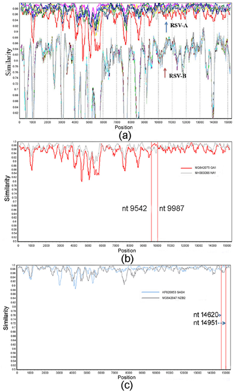
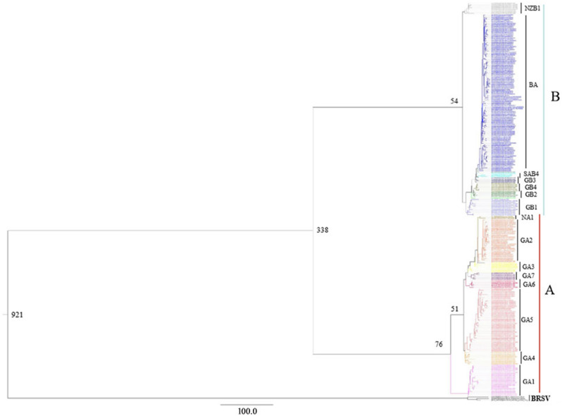
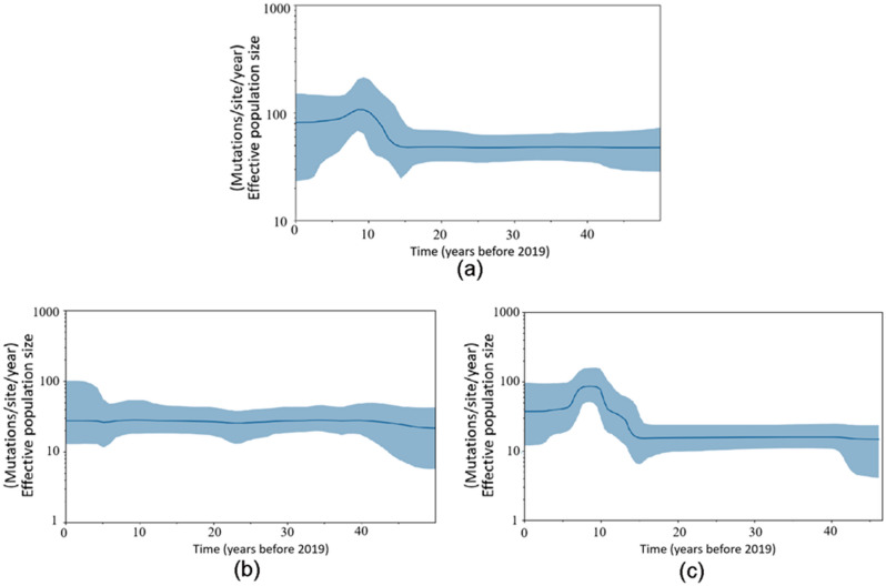
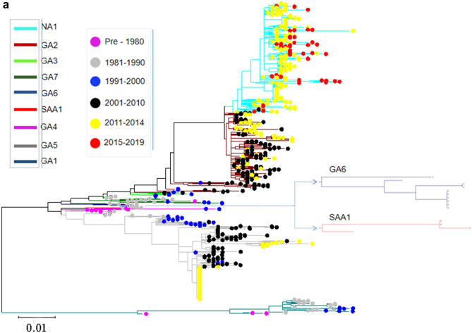
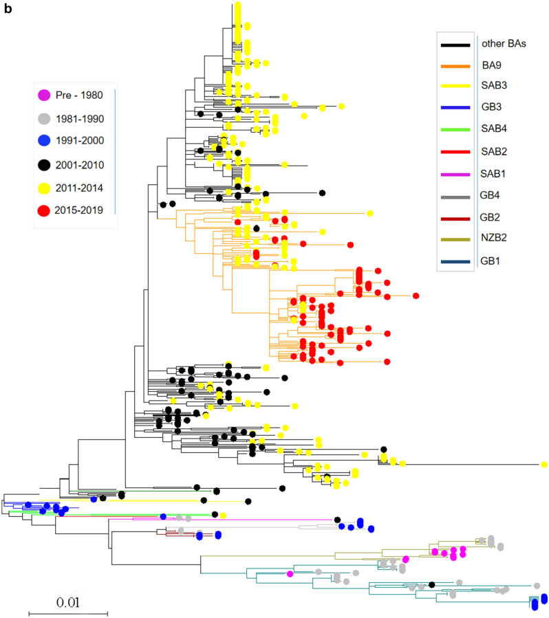
Human respiratory syncytial viruses (RSVs) are classified into two major groups (A and B) based on antigenic differences in the G glycoprotein. To investigate circulating characteristics and phylodynamic history of RSV, we analyzed the genetic variability and evolutionary pattern of RSVs from 1977 to 2019 in this study. The results revealed that there was no recombination event of intergroup. Single nucleotide polymorphisms (SNPs) were observed through the genome with the highest occurrence rate in the G gene. Five and six sites in G protein of RSV-A and RSV-B, respectively, were further identified with a strong positive selection. The mean evolutionary rates for RSV-A and -B were estimated to be 1.48 × 10⁻³ and 1.92 × 10⁻³ nucleotide substitutions/site/year, respectively. The Bayesian skyline plot showed a constant population size of RSV-A and a sharp expansion of population size of RSV-B since 2005, and an obvious decrease 5 years later, then became stable again. The total population size of RSVs showed a similar tendency to that of RSV-B. Time-scaled phylogeny suggested a temporal specificity of the RSV-genotypes. Monitoring nucleotide changes and analyzing evolution pattern for RSVs could give valuable insights for vaccine and therapy strategies against RSV infection.


Conflict of interest statement

The authors declare no competing interests.

Figures

Figure 1
Figure 1
Recombination analysis in RSVs by using representative subtypes/genotypes. (a) RSV-A and RSV-B: no recombination event was observed; (b) RSV-A and (c) RSV-B: potential recombination breakpoints in the intragroups were marked in red lines and the positions were annotated.
Figure 2
Figure 2
Bayesian Markov Chain Monte Carlo tree of the complete G genes of RSV-A, RSV-B and BRSV. The estimated time of the MRCA of the major nodes and the names of different viral clades were marked in the graph. The results showed that RSV-A and RSV-B diverged from their common ancestor 338 years ago.
Figure 3
Figure 3
Population dynamics of the G gene for the RSV population using Bayesian skyline plot analyses. (a) both the RSV-A and RSV-B strains; (b) RSV-A; (c) RSV-B. The thick solid lines indicate mean effective population sizes; blue shadings stand for 95% HPD.
Figure 4
Figure 4
Temporal distribution of evolutionary clusters in the unrooted phylogenetic tree based on the RSV G gene sequence alignments using maximum likelihood method. (a) RSV-A: the invisible GA6 and SAA1 were further enlarged on the right side of the figure; (b) RSV-B. Dots with different color indicate sequences from different time, and lines with different color indicate different subtypes.
Figure 4
Figure 4
Temporal distribution of evolutionary clusters in the unrooted phylogenetic tree based on the RSV G gene sequence alignments using maximum likelihood method. (a) RSV-A: the invisible GA6 and SAA1 were further enlarged on the right side of the figure; (b) RSV-B. Dots with different color indicate sequences from different time, and lines with different color indicate different subtypes.

References

    1. Shi T, et al. Global, regional, and national disease burden estimates of acute lower respiratory infections due to respiratory syncytial virus in young children in 2015: A systematic review and modelling study. Lancet. 2017;390:946–958. doi: 10.1016/S0140-6736(17)30938-8. - DOI - PMC - PubMed
    1. Anderson LJ, et al. Antigenic characterization of respiratory syncytial virus strains with monoclonal antibodies. J. Infect. Dis. 1985;151:626–633. doi: 10.1093/infdis/151.4.626. - DOI - PubMed
    1. Mufson MA, Orvell C, Rafnar B, Norrby E. Two distinct subtypes of human respiratory syncytial virus. J. Gen. Virol. 1985;66(Pt 10):2111–2124. doi: 10.1099/0022-1317-66-10-2111. - DOI - PubMed
    1. Johnson PR, Spriggs MK, Olmsted RA, Collins PL. The G glycoprotein of human respiratory syncytial viruses of subgroups A and B: Extensive sequence divergence between antigenically related proteins. Proc. Natl. Acad. Sci. USA. 1987;84:5625–5629. doi: 10.1073/pnas.84.16.5625. - DOI - PMC - PubMed
    1. Martinez I, Dopazo J, Melero JA. Antigenic structure of the human respiratory syncytial virus G glycoprotein and relevance of hypermutation events for the generation of antigenic variants. J. Gen. Virol. 1997;78(Pt 10):2419–2429. doi: 10.1099/0022-1317-78-10-2419. - DOI - PubMed

Publication types