

Images in Primary Care Medicine: Point-of-Care Ultrasound in Gout
- PMID: 34155462
- PMCID: PMC8211301
- DOI: 10.7759/cureus.15096
Images in Primary Care Medicine: Point-of-Care Ultrasound in Gout
Abstract
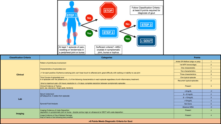
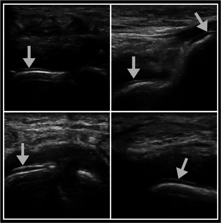
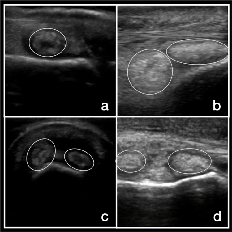
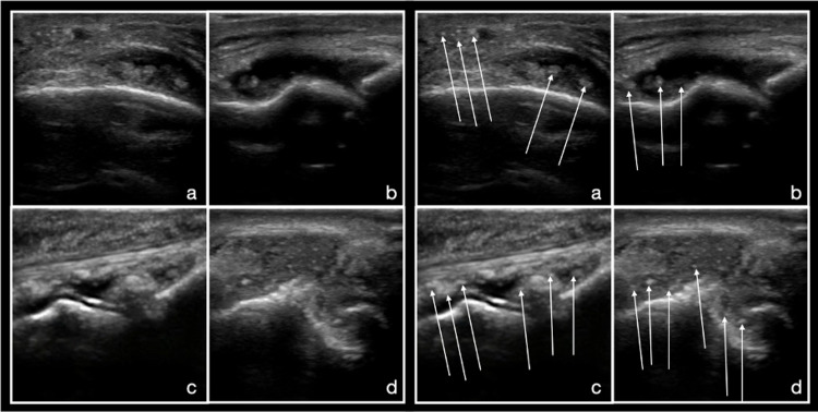
Gout is the most common crystal arthropathy and is frequently diagnosed and managed by primary care physicians. Point-of-care ultrasound (POCUS) is a valuable tool to aid in the diagnosis of gout via the identification of the double contour sign, aggregates of crystals, tophi, and erosions. In addition, POCUS can aid in the management of gout by recognizing early signs of gout, monitoring the effectiveness of urate-lowering therapy, and guiding aspiration and corticosteroid injection.
Keywords: crystal arthropathy; gout crystals; point-of-care-ultrasound; primary care medicine; ultrasound-guided.
Copyright © 2021, Espejo et al.
Conflict of interest statement
The authors have declared that no competing interests exist.
Figures
Similar articles
-
Gouty arthropathy: Review of clinico-pathologic and imaging features.J Med Imaging Radiat Oncol. 2016 Feb;60(1):9-20. doi: 10.1111/1754-9485.12356. Epub 2015 Oct 5. J Med Imaging Radiat Oncol. 2016. PMID: 26439321 Review.
-
Ultrasound and clinical features of hip involvement in patients with gout.Joint Bone Spine. 2019 Oct;86(5):633-636. doi: 10.1016/j.jbspin.2019.01.027. Epub 2019 Feb 16. Joint Bone Spine. 2019. PMID: 30779966
-
The popliteal groove region: A new target for the detection of monosodium urate crystal deposits in patients with gout. An ultrasound study.Joint Bone Spine. 2019 Jan;86(1):89-94. doi: 10.1016/j.jbspin.2018.06.008. Epub 2018 Jul 17. Joint Bone Spine. 2019. PMID: 30025961
-
Combining Hyperechoic Aggregates and the Double-Contour Sign Increases the Sensitivity of Sonography for Detection of Monosodium Urate Deposits in Gout.J Ultrasound Med. 2017 May;36(5):935-940. doi: 10.7863/ultra.16.03046. Epub 2017 Feb 27. J Ultrasound Med. 2017. PMID: 28240795
-
What do I need to know about gout?J Fam Pract. 2010 Jun;59(6 Suppl):S1-8. J Fam Pract. 2010. PMID: 20544070 Review.
References
-
- Comorbidities in patients with crystal diseases and hyperuricemia. Sattui SE, Singh JA, Gaffo AL. https://www.ncbi.nlm.nih.gov/pmc/articles/PMC4159668/ Rheum Dis Clin North Am. 2014;40:251–278. - PMC - PubMed
-
- Global epidemiology of gout: prevalence, incidence and risk factors. Kuo CF, Grainge MJ, Zhang W, Doherty M. Nat Rev Rheumatol. 2015;11:649–662. - PubMed
-
- Ambulatory resource utilization and cost for gout in United States. Li C, Martin BC, Cummins DF, Andrews LM, Frech-Tamas F, Yadao AM. https://www.researchgate.net/publication/286605969_Ambulatory_resource_u... Am J Pharm Benefits. 2013;5:46–54.
-
- Diagnosis, treatment, and prevention of gout. Hainer BL, Matheson E, Wilkes RT. https://www.aafp.org/afp/2014/1215/p831.html Am Fam Physician. 2014;15:831–836. - PubMed
-
- Gout and hyperuricaemia in the USA: prevalence and trends. Singh G, Lingala B, Mithal A. Rheumatology (Oxford) 2019;58:2177–2180. - PubMed
Publication types
LinkOut - more resources
Full Text Sources