

Review
doi: 10.3346/jkms.2021.36.e177.
Osteonecrosis of the Femoral Head: an Updated Review of ARCO on Pathogenesis, Staging and Treatment
Affiliations
- PMID: 34155839
- PMCID: PMC8216992
- DOI: 10.3346/jkms.2021.36.e177
Item in Clipboard
Review
Osteonecrosis of the Femoral Head: an Updated Review of ARCO on Pathogenesis, Staging and Treatment
J Korean Med Sci.
.
Abstract
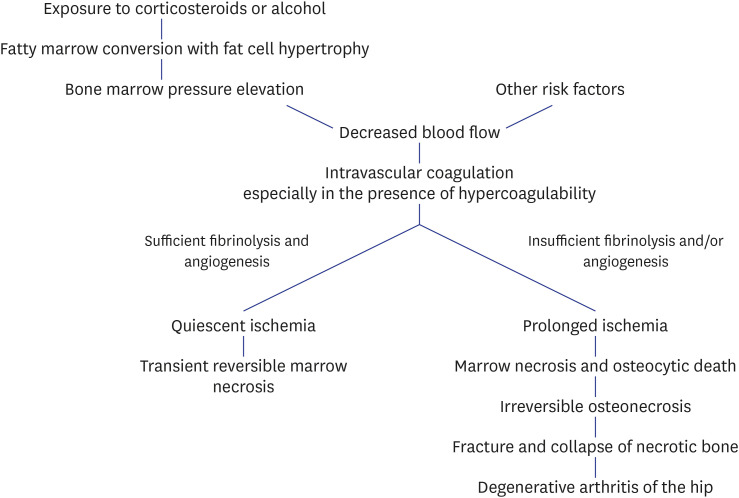
Non-traumatic osteonecrosis of the femoral head (ONFH) usually affects adults younger than 50 years and frequently leads to femoral head collapse and subsequent arthritis of the hip. It is becoming more prevalent along with increasing use of corticosteroids for the adjuvant therapy of leukemia and other myelogenous diseases as well as management of organ transplantation. This review updated knowledge on the pathogenesis, classification criteria, staging system, and treatment of ONFH.
Keywords: Avascular Necrosis; Femoral Head; Hip; Osteonecrosis.
© 2021 The Korean Academy of Medical Sciences.
Conflict of interest statement
The authors have no potential conflicts of interest to disclose.
Figures
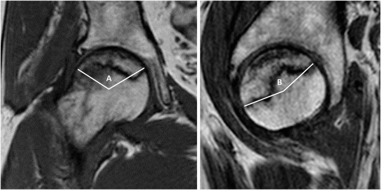
MR = magnetic resonance.
References
-
- Dubois EL, Cozen L. Avascular (aseptic) bone necrosis associated with systemic lupus erythematosus. JAMA. 1960;174(8):966–971. - PubMed
-
- Phemister DB. Changes in bones and joints resulting from interruption of circulation. I. General consideration and changes resulting from injury. Arch Surg. 1940;41(2):436.
-
- Mont MA, Cherian JJ, Sierra RJ, Jones LC, Lieberman JR. Nontraumatic osteonecrosis of the femoral head: Where do we stand today? A ten-year update. J Bone Joint Surg Am. 2015;97(19):1604–1627. - PubMed
-
- Kang JS, Park S, Song JH, Jung YY, Cho MR, Rhyu KH. Prevalence of osteonecrosis of the femoral head: a nationwide epidemiologic analysis in Korea. J Arthroplasty. 2009;24(8):1178–1183. - PubMed
Publication types
MeSH terms
Substances
LinkOut - more resources
Full Text Sources
Medical
