Observational Study
. 2021 Jul;69(7):1670-1692.
doi: 10.4103/ijo.IJO_1565_21.

Epidemiology, clinical profile, management, and outcome of COVID-19-associated rhino-orbital-cerebral mucormycosis in 2826 patients in India - Collaborative OPAI-IJO Study on Mucormycosis in COVID-19 (COSMIC), Report 1

Mrittika Sen  1 Santosh G Honavar  1 Rolika Bansal  1 Sabyasachi Sengupta  2 Raksha Rao  3 Usha Kim  4 Mukesh Sharma  5 Mahipal Sachdev  6 Ashok K Grover  7 Abhidnya Surve  8 Abhishek Budharapu  9 Abhishek K Ramadhin  10 Abhishek Kumar Tripathi  11 Adit Gupta  12 Aditya Bhargava  13 Animesh Sahu  14 Anjali Khairnar  15 Anju Kochar  16 Ankita Madhavani  17 Ankur K Shrivastava  18 Anuja K Desai  19 Anujeet Paul  20 Anuradha Ayyar  21 Aparna Bhatnagar  22 Aparna Singhal  23 Archana Sunil Nikose  24 Arun Bhargava  14 Arvind L Tenagi  25 Ashish Kamble  26 Ashiyana Nariani  27 Bhavin Patel  28 Bibbhuti Kashyap  29 Bodhraj Dhawan  30 Busaraben Vohra  31 Charuta Mandke  32 Chinmayee Thrishulamurthy  33 Chitra Sambare  34 Deepayan Sarkar  35 Devanshi Shirishbhai Mankad  17 Dhwani Maheshwari  36 Dilip Lalwani  37 Dipti Kanani  17 Diti Patel  31 Fairooz P Manjandavida  38 Frenali Godhani  39 Garima Amol Agarwal  40 Gayatri Ravulaparthi  41 Gondhi Vijay Shilpa  42 Gunjan Deshpande  43 Hansa Thakkar  40 Hardik Shah  44 Hare Ram Ojha  45 Harsha Jani  46 Jyoti Gontia  47 Jyotika P Mishrikotkar  48 Kamalpreet Likhari  49 Kamini Prajapati  40 Kavita Porwal  50 Kirthi Koka  51 Kulveer Singh Dharawat  52 Lakshmi B Ramamurthy  53 Mainak Bhattacharyya  54 Manorama Saini  23 Marem C Christy  1 Mausumi Das  20 Maya Hada  52 Mehul Panchal  55 Modini Pandharpurkar  42 Mohammad Osman Ali  42 Mukesh Porwal  56 Nagaraju Gangashetappa  33 Neelima Mehrotra  57 Neha Bijlani  58 Nidhi Gajendragadkar  29 Nitin M Nagarkar  59 Palak Modi  40 Parveen Rewri  23 Piyushi Sao  60 Prajakta Salunkhe Patil  61 Pramod Giri  43 Priti Kapadia  62 Priti Yadav  47 Purvi Bhagat  40 Ragini Parekh  63 Rajashekhar Dyaberi  53 Rajender Singh Chauhan  64 Rajwinder Kaur  65 Ram Kishan Duvesh  66 Ramesh Murthy  67 Ravi Varma Dandu  68 Ravija Kathiara  69 Renu Beri  70 Rinal Pandit  71 Rita Hepsi Rani  72 Roshmi Gupta  3 Ruchi Pherwani  73 Rujuta Sapkal  69 Rupa Mehta  70 Sameeksha Tadepalli  74 Samra Fatima  42 Sandeep Karmarkar  75 Sandeep Suresh Patil  76 Sanjana Shah  31 Sankit Shah  77 Sapan Shah  19 Sarika Dubey  52 Saurin Gandhi  78 Savitha Kanakpur  53 Shalini Mohan  79 Sharad Bhomaj  80 Sheela Kerkar  27 Shivani Jariwala  62 Shivati Sahu  47 Shruthi Tara  81 Shruti Kochar Maru  50 Shubha Jhavar  82 Shubhda Sharma  83 Shweta Gupta  83 Shwetha Kumari  84 Sima Das  85 Smita Menon  27 Snehal Burkule  86 Sonam Poonam Nisar  51 Subashini Kaliaperumal  83 Subramanya Rao  33 Sudipto Pakrasi  83 Sujatha Rathod  33 Sunil G Biradar  60 Suresh Kumar  87 Susheen Dutt  88 Svati Bansal  83 Swati Amulbhai Ravani  40 Sweta Lohiya  89 Syed Wajahat Ali Rizvi  90 Tanmay Gokhale  91 Tatyarao P Lahane  63 Tejaswini Vukkadala  92 Triveni Grover  93 Trupti Bhesaniya  62 Urmil Chawla  64 Usha Singh  74 Vaishali L Une  15 Varsha Nandedkar  82 Venkata Subramaniam  94 Vidya Eswaran  84 Vidya Nair Chaudhry  95 Viji Rangarajan  96 Vipin Dehane  97 Vivek M Sahasrabudhe  86 Yarra Sowjanya  98 Yashaswini Tupkary  99 Yogita Phadke  69 members of the Collaborative OPAI-IJO Study on Mucormycosis in COVID-19 (COSMIC) Study Group
Affiliations
Observational Study

Epidemiology, clinical profile, management, and outcome of COVID-19-associated rhino-orbital-cerebral mucormycosis in 2826 patients in India - Collaborative OPAI-IJO Study on Mucormycosis in COVID-19 (COSMIC), Report 1

Mrittika Sen et al. Indian J Ophthalmol. 2021 Jul.

Abstract

Purpose: COVID-19-associated rhino-orbital-cerebral mucormycosis (ROCM) has reached epidemic proportion during India's second wave of COVID-19 pandemic, with several risk factors being implicated in its pathogenesis. This study aimed to determine the patient demographics, risk factors including comorbidities, and medications used to treat COVID-19, presenting symptoms and signs, and the outcome of management.

Methods: This was a retrospective, observational study of patients with COVID-19-associated ROCM managed or co-managed by ophthalmologists in India from January 1, 2020 to May 26, 2021.

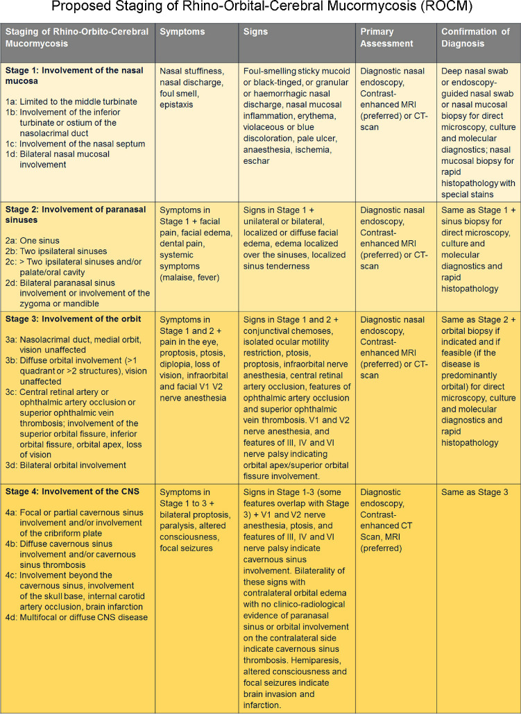
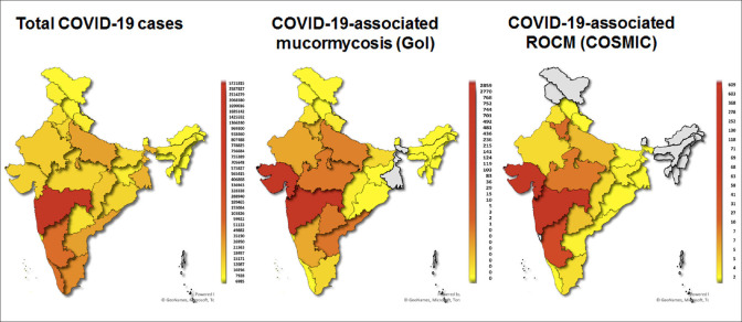
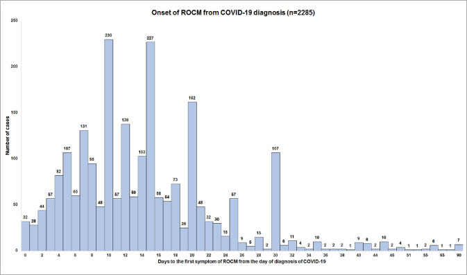
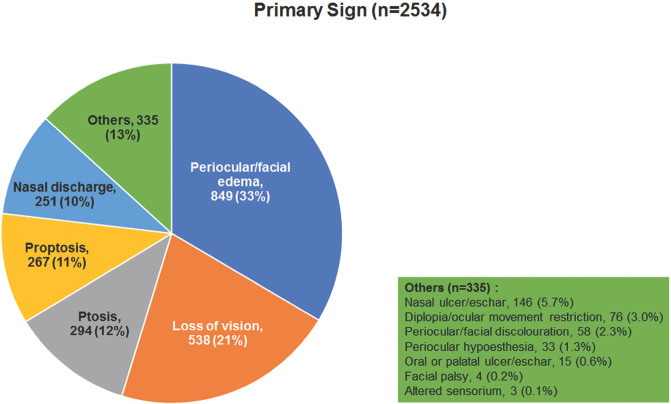
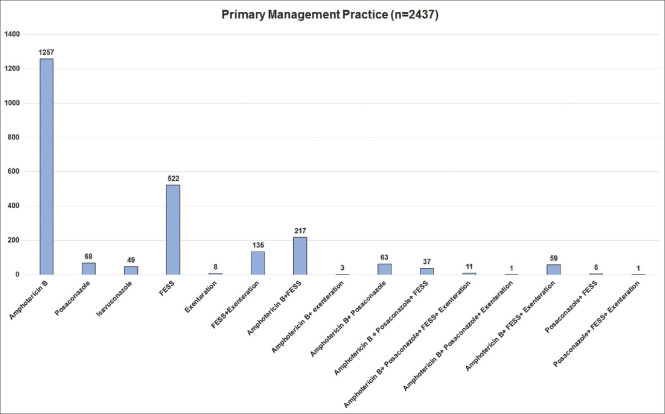
Results: Of the 2826 patients, the states of Gujarat (22%) and Maharashtra (21%) reported the highest number of ROCM. The mean age of patients was 51.9 years with a male preponderance (71%). While 57% of the patients needed oxygen support for COVID-19 infection, 87% of the patients were treated with corticosteroids, (21% for > 10 days). Diabetes mellitus (DM) was present in 78% of all patients. Most of the cases showed onset of symptoms of ROCM between day 10 and day 15 from the diagnosis of COVID-19, 56% developed within 14 days after COVID-19 diagnosis, while 44% had delayed onset beyond 14 days. Orbit was involved in 72% of patients, with stage 3c forming the bulk (27%). Overall treatment included intravenous amphotericin B in 73%, functional endoscopic sinus surgery (FESS)/paranasal sinus (PNS) debridement in 56%, orbital exenteration in 15%, and both FESS/PNS debridement and orbital exenteration in 17%. Intraorbital injection of amphotericin B was administered in 22%. At final follow-up, mortality was 14%. Disease stage >3b had poorer prognosis. Paranasal sinus debridement and orbital exenteration reduced the mortality rate from 52% to 39% in patients with stage 4 disease with intracranial extension (p < 0.05).

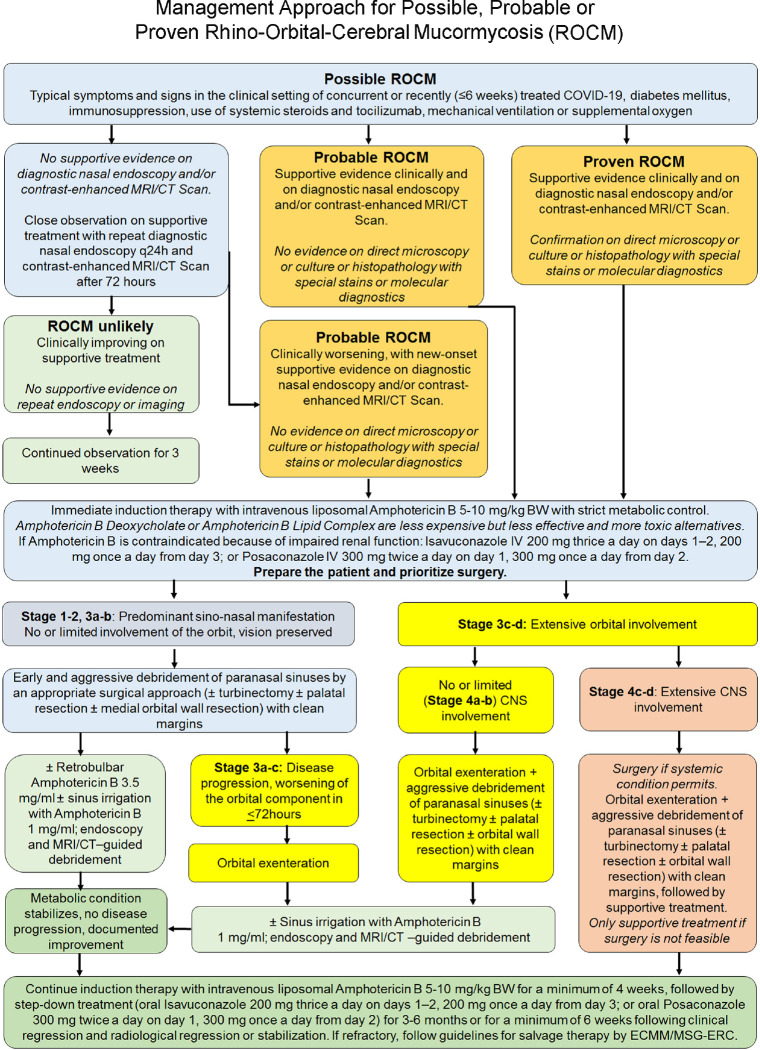
Conclusion: : Corticosteroids and DM are the most important predisposing factors in the development of COVID-19-associated ROCM. COVID-19 patients must be followed up beyond recovery. Awareness of red flag symptoms and signs, high index of clinical suspicion, prompt diagnosis, and early initiation of treatment with amphotericin B, aggressive surgical debridement of the PNS, and orbital exenteration, where indicated, are essential for successful outcome.

Keywords: COVID-19; COVID-19-associated ROCM; Corticosteroids; diabetes mellitus; mucormycosis; orbital exenteration; paransal sinus debridement; rhino-orbital-cerebral mucormycosis; staging of rhino-orbital-cerebral mucormycosis.


Conflict of interest statement

There are no conflicts of interest.

Figures

Figure 1
Figure 1
Proposed staging system for COVID-19 associated rhino-orbital-cerebral mucormycosis (Reproduced with permission from Honavar SG. Code Mucor: Guidelines for the Diagnosis, Staging and Management of Rhino-Orbito-Cerebral Mucormycosis in the Setting of COVID-19. Indian Journal of Ophthalmology. 2021;69:1361-5)[6]
Figure 2
Figure 2
State-wise distribution of COVID-19-positive cases in India (as on June 2, 2021) (left) compared with state-wise hot-spots of COVID-19-associated rhino-orbital-cerebral mucormycosis (ROCM) cases, national data on COVID-19-associated mucormycosis (centre)[7] and COSMIC group data (right). Gujarat, Madhya Pradesh, Haryana and Telangana seem to have disproportionately more ROCM cases, while Kerala, Tamil Nadu, Andhra Pradesh and West Bengal seem to have relatively less ROCM cases as compared to the cases of COVID-19 being reported. (The COSMIC data includes only those cases that have been submitted for the study and may not be representative of the actual incidence).
Figure 3
Figure 3
Frequency of cases with onset of rhino-orbital-cerebral mucormycosis symptoms from the day of diagnosis of COVID-19 (Day 0)
Figure 4
Figure 4
Frequency of primary symptoms in patients with COVID-19-associated rhino-orbital-cerebral mucormycosis
Figure 5
Figure 5
Frequency of primary signs in patients with COVID-19-associated rhino-orbital-cerebral mucormycosis
Figure 6
Figure 6
Stage 1 rhino-orbital-cerebral mucormycosis. Nasal endoscopy pictures showing (a) Stage 1a: Isolated involvement of the left middle turbinate (white arrow) (b) Stage 1b: Involvement of the left inferior turbinate (white arrow) (c) Stage 1c: Involvement of the left nasal septum (white arrow) (d, e) Stage 1d: Bilateral nasal mucosal involvement (right and left side respectively) (white arrow) (f) Stage 2c rhino-orbital-cerebral mucormycosis. Coronal MRI (Post contrast T1 image with fat saturation) orbit and paranasal sinuses showing the non-enhancing inferior and middle turbinate – the “black turbinate sign” (red arrow). The “black turbinate sign” is one of the earliest MRI signs of rhino-orbital-cerebral mucormycosis, and can be detected in stage 1. (Endoscopy images provided by Sandeep Karmarkar, MRI image provided by Ravi Varma)
Figure 7
Figure 7
Stage 2a rhino-orbital-cerebral mucormycosis (a) Clinical photograph showing left periocular edema with mild ptosis, conjunctival congestion and chemosis. (b) Axial MRI (T2 with fat saturation) showing mucosal thickening in the left ethmoid sinus. Stage 2b rhino-orbitalcerebral mucormycosis (c) Coronal MRI (T2) of the orbit and paranasal sinuses showing mucosal thickening in the right maxillary and ethmoid sinuses (d). Coronal post-contrast (T1) MRI showing enhancement of the mucosa limited to the ipsilateral maxillary and ethmoid sinuses (Clinical image provided by Chinmayee T, MRI images by Ravi Varma)
Figure 8
Figure 8
Stage 2c rhino-orbital-cerebral mucormycosis (a, b) Clinical photographs showing palatal involvement with a visible black eschar (c) Coronal MRI (T1) and (d) Coronal MRI (T2) of the orbit and paranasal sinuses showing mucosal thickening in >2 ipsilateral sinuses along with palatal involvement (red arrow) (e) Endoscopy picture showing necrosed tissue in left sphenoid sinus (white arrow) and left maxillary sinus (yellow arrow). (Clinical images provided by Chinmayee T, MRI images by Ravi Varma, endoscopy image by Sandeep Karmarkar)
Figure 9
Figure 9
Stage 2d rhino-orbital-cerebral mucormycosis (a) Axial MRI (T2) and Coronal post contrast T1 with fat saturation (b) of the orbit and paranasal sinuses showing mucosal thickening and enhancement in bilateral ethmoid and maxillary sinuses (c) Endoscopic picture showing right frontal sinus involvement with necrotic mucosa (white arrow) along with (d) left sided involvement of sphenoid (white arrow) and maxillary sinus (yellow arrow). (Endoscopy images provided by Sandeep Karmarkar)
Figure 10
Figure 10
Stage 3a rhino-orbital-cerebral mucormycosis (a) Clinical picture showing left periocular edema, ptosis and proptosis (b) Endoscopy picture showing necrosed left periorbita (white arrow) (c, d) Coronal and axial T2 weighted MR images showing involvement of the right nasolacrimal duct (red arrows), extending into the medial orbit. (Endoscopy image provided by Sandeep Karmarkar, MRI images by Ravi Varma)
Figure 11
Figure 11
Stage 3b rhino-orbital-cerebral mucormycosis (a) Clinical pictures showing the right eye proptosis with restriction of ocular movements in all gazes. (b) Axial contrast-enhanced MRI (T1) of the orbit, paranasal sinuses, and brain showing involvement of the medial orbit and abnormal intensity of the orbital fat in the posterior orbit along with involvement of the right ethmoid sinus
Figure 12
Figure 12
Stage 3c rhino-orbital-cerebral mucormycosis (a) Clinical picture showing right eye ptosis, periocular edema and discolouration (b) severe conjunctival congestion and chemosis (c) Axial MRI (T2) and axial post contrast (T1) with fat saturation showing right ethmoid sinus and diffuse orbital involvement extending to the orbital apex (MRI images provided by Ravi Varma)
Figure 13
Figure 13
Stage 3d rhino-orbital-cerebral mucormycosis (a) Axial MRI (T2) and (b) contrast enhanced (T1) of the orbit and paranasal sinuses showing bilateral orbital apical involvement, more extensive on the right side. (Images provided by Ravi Varma)
Figure 14
Figure 14
Stage 4a rhino-orbital-cerebral mucormycosis (a) Clinical photograph showing restriction of ocular movements in all gazes in the left eye. (b) Axial MRI (T1) of the orbit and brain showing diffuse orbital involvement along with focal cavernous sinus involvement (red arrow) (c) Endoscopy picture showing eroded left cribriform plate (white arrow) with dural defect (yellow arrow) (Endoscopy image provided by Sandeep Karmarkar)
Figure 15
Figure 15
Stage 4b rhino-orbital-cerebral mucormycosis (a) Clinical picture showing no significant ocular manifestation, however, (b) Axial and (c) coronal T1 MRI with fat saturation of the orbit and paranasal sinuses and brain showed involvement of the sphenoid sinus with extension into the cavernous sinus. (MRI images provided by Ravi Varma)
Figure 16
Figure 16
Stage 4c rhino-orbital-cerebral mucormycosis (a) Primary gaze pictures showing ptosis of the right eye along with periocular edema (b) MR angiogram showing left internal carotid artery occlusion (MR Angiography image provided by Chinmayee T)
Figure 17
Figure 17
Stage 4d rhino-orbital-cerebral mucormycosis (a, b, c) MRI (Diffusion imaging) showing multifocal hyperintense areas indicating diffuse cerebral parenchymal involvement. (d) Coronal post contrast T1 MRI (T1) showing temporal lobe abscess (red arrow) (e) Endoscopic picture showing right sided temporal abscess cavity (yellow arrow). (MRI image (d) and endoscopic image provided by Sandeep Karmarkar)
Figure 18
Figure 18
Primary management of patients with COVID-19-associated rhino-orbital-cerebral mucormycosis
Figure 19
Figure 19
Proposed management algorithm for rhino-orbital-cerebral mucormycosis (Reproduced with permission from Honavar SG. Code Mucor: Guidelines for the Diagnosis, Staging and Management of Rhino-Orbito-Cerebral Mucormycosis in the Setting of COVID-19. Indian Journal of Ophthalmology. 2021 Jun;69:1361-5)[6]

References

    1. [Last accessed on 2021 May 31]. Available from: https://www.nytimes.com/interactive/2021/world/india-covid-cases.html .
    1. Singh AK, Singh R, Joshi SR, Misra A. Mucormycosis in COVID-19:A systematic review of cases reported worldwide and in India. Diabetes Metab Syndr. 2021 doi:10.1016/j.dsx. 2021.05.019. - PMC - PubMed
    1. Sen M, Lahane S, Lahane TP, Parekh R, Honavar SG. Mucor in a viral land:A tale of two pathogens. Indian J Ophthalmol. 2021;69:244–52. - PMC - PubMed
    1. Ravani SA, Agrawal GA, Leuva PA, Modi PH, Amin KD. Rise of the phoenix:Mucormycosis in COVID-19 times. Indian J Ophthalmol. 2021;69:1563–8. - PMC - PubMed
    1. Sarkar S, Gokhale T, Choudhury SS, Deb AK. COVID-19 and orbital mucormycosis. Indian J Ophthalmol. 2021;69:1002–4. - PMC - PubMed

Publication types

Substances