Review
. 2021 Aug;8(8):1709-1719.
doi: 10.1002/acn3.51401. Epub 2021 Jun 22.

Mechanistic underpinning of an inside-out concept for autoimmunity in multiple sclerosis

Affiliations
Review

Mechanistic underpinning of an inside-out concept for autoimmunity in multiple sclerosis

Bert A 't Hart et al. Ann Clin Transl Neurol. 2021 Aug.

Abstract

The neuroinflammatory disease multiple sclerosis is driven by autoimmune pathology in the central nervous system. However, the trigger of the autoimmune pathogenic process is unknown. MS models in immunologically naïve, specific-pathogen-free bred rodents support an exogenous trigger, such as an infection. The validity of this outside-in pathogenic concept for MS has been frequently challenged by the difficulty to translate pathogenic concepts developed in these models into effective therapies for the MS patient. Studies in well-validated non-human primate multiple sclerosis models where, just like in humans, the autoimmune pathogenic process develops from an experienced immune system trained by prior infections, rather support an endogenous trigger. Data reviewed here corroborate the validity of this inside-out pathogenic concept for multiple sclerosis. They also provide a plausible sequence of events reminiscent of Wilkin's primary lesion theory: (i) that autoimmunity is a physiological response of the immune system against excess antigen turnover in diseased tissue (the primary lesion) and (ii) that individuals developing autoimmune disease are (genetically predisposed) high responders against critical antigens. Data obtained in multiple sclerosis brains reveal the presence in normally appearing white matter of myelinated axons where myelin sheaths have locally dissociated from their enwrapped axon (i.e., blistering). The ensuing disintegration of axon-myelin units potentially causes the excess systemic release of post-translationally modified myelin. Data obtained in a unique primate multiple sclerosis model revealed a core pathogenic role of T cells present in the normal repertoire, which hyper-react to post-translationally modified (citrullinated) myelin-oligodendrocyte glycoprotein and evoke clinical and pathological aspects of multiple sclerosis.


Conflict of interest statement

No disclosures relevant to the publication are reported.

Figures
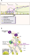
Figure 1
Figure 1
Clinical course of MS (A) and the targets of the autoimmune attack (B). (A) In the majority of MS patients (>80%) their disease starts with alternating episodes of neurological disability (relapse) and recovery (remission); this phase is indicated as relapsing‐remitting MS (RRMS). However, prior to symptom diagnosis, focal inflammation of white matter can be visualized with MRI; this is, therefore, indicated as pre‐symptomatic MS. In about 60% of patients with RRMS, conversion to progressive worsening of symptoms with decreasing remission occurs; this is secondary progressive (SP) MS. Patients with primary progressive MS (±15%) seem to have skipped the RR phase; their disease is progressive from the onset. Intriguingly, MRI‐detectable white matter inflammation is substantially lower in PMS than in RRMS. (B) Depicted is one oligodendrocyte that forms myelin sheaths around four different axons. The autoimmune attack on myelin sheaths (outside–in) comprises binding of autoantibody to a surface‐exposed antigen (MOG?). This complex is bound by a macrophage, eliciting antibody‐dependent cytotoxicity (ADCC), or by complement factors, eliciting complement‐dependent cytotoxicity (CDC). Oligodendrocytes can be directly attacked by CD8+ T cells, which react to MHC‐mediated presentation of myelin antigens. Injury by primary oligodendrogliopathy occurs independent of an immune attack and may lead to myelin sheath disintegration starting at the inner myelin lamellae (panel B has been reproduced from: 't Hart et al. eBiomedicine 2021, in press).
Figure 2
Figure 2
Swelling types of the axon–myelin unit. Depicted in 1a are the three types of swellings of myelinated axons identified in MS normal‐appearing white matter, placed in a putative sequence: normal axon–myelin unit => myelin detachment from axon (blister) => swelling of the demyelinated axon segment (bleb) => degeneration of axon and myelin resulting in the release of myelin debris (1b). In areas enriched with such swellings, myelin proteins are post‐translationally modified by citrullination, that is, the enzymatic substitution of positively charged arginine for neutrally charged citrulline. 2b. Myelin debris containing citrullinated MOG are taken up by EBV‐infected B cells and processed intracellularly in phagolysosomal compartments. Presentation of strongly immunogenic citrullinated MOG results in autoreactive T cell activation.
Figure 3
Figure 3
Formation of self‐aggregating structures of MOG34‐56 and citrullinated derivatives in overnight incubation with or without human EBV‐infected B cell (EBV‐BC) line. Native MOG34‐56 (row A) and the citrullinated versions MOGcitr41,46 (row B) and MOGcitr46,52 (row C) were fed to EBV‐BC. The peptides were labeled with purple fluorochrome via click chemistry. The cells were stained with DAPI (blue) to visualize nuclei and with anti‐LC3 (autophagosomes) or anti‐HLA‐DR (B cell membrane antigen) mAb (both green). The figure shows low background LC3 staining of EBV‐BCL incubated without peptide (A1). Feeding MOG34‐56 increases LC3 staining (A3,4) indicating upregulation of autophagy. The peptide appears in spherical structures (A3,4 purple) of varying size. Citrullinated MOGcitr41,46 peptide incubated without cells spontaneously forms medium‐sized aggregates (B2). When incubated with EBV‐BC at a low dose similar spherical structures as the native peptide (B3). Containment in an MHC class II+ staining capsule (B5) suggests that the spheres originate from the EBV‐BC. Incubates of EBV‐BC with high dose MOGcitr41,46 (B4) contain large peptide aggregates and virtually no B cells, indicating that these may have been killed, as confirmed in reference. MOGcitr46,52 peptide incubated without EBV‐BC (C2) or with EBV‐BC (C3,4) spontaneously forms much larger aggregates than in B2 and at a much higher rate (see Ref. [57]), but spherical structures were not found.

References

    1. Starr TK, Jameson SC, Hogquist KA. Positive and negative selection of T cells. Annu Rev Immunol 2003;21:139–176. - PubMed
    1. Sercarz EE, Lehmann PV, Ametani A, et al. Dominance and crypticity of T cell antigenic determinants. Annu Rev Immunol 1993;11:729–766. - PubMed
    1. Serre L, Girard M, Ramadan A, et al. Thymic‐specific serine protease limits central tolerance and exacerbates experimental autoimmune encephalomyelitis. J Immunol 2017;199:3748–3756. - PubMed
    1. Meinl E, Weber F, Drexler K, et al. Myelin basic protein‐specific T lymphocyte repertoire in multiple sclerosis. Complexity of the response and dominance of nested epitopes due to recruitment of multiple T cell clones. J Clin Invest 1993;92:2633–2643. - PMC - PubMed
    1. ’t Hart BA, Gran B, Weissert R. EAE: imperfect but useful models of multiple sclerosis. Trends Mol Med 2011;17:119–125. - PubMed