

GHRH secretion from a pancreatic neuroendocrine tumor causing gigantism in a patient with MEN1
- PMID: 34156350
- PMCID: PMC8240703
- DOI: 10.1530/EDM-20-0208
GHRH secretion from a pancreatic neuroendocrine tumor causing gigantism in a patient with MEN1
Abstract
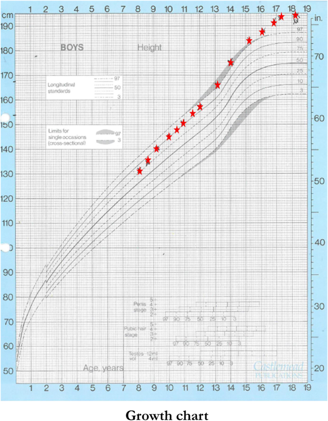
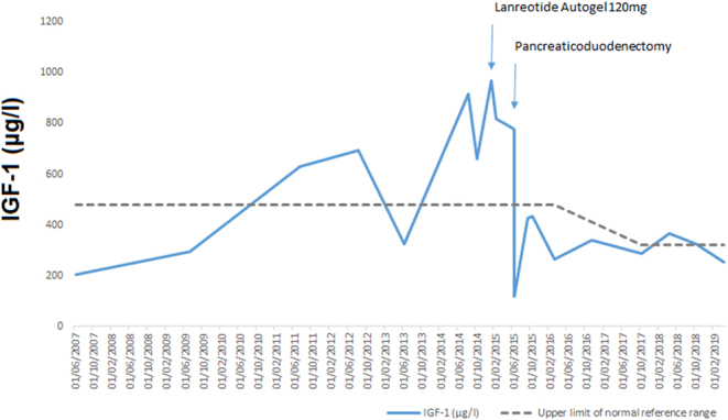
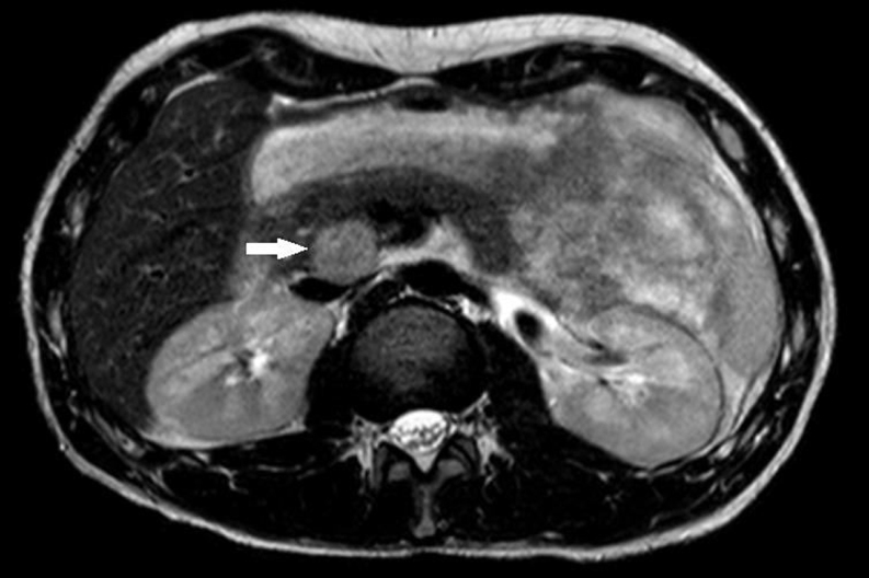
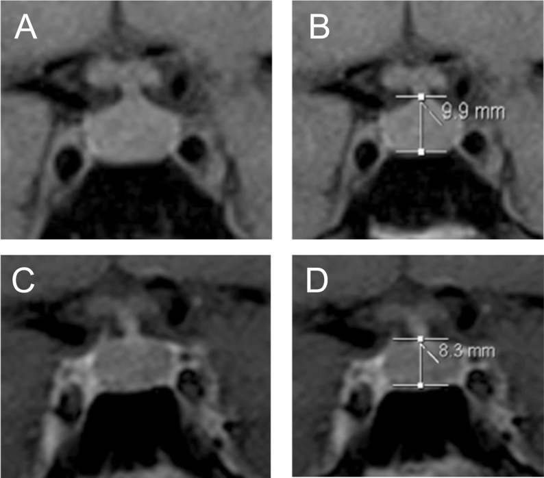
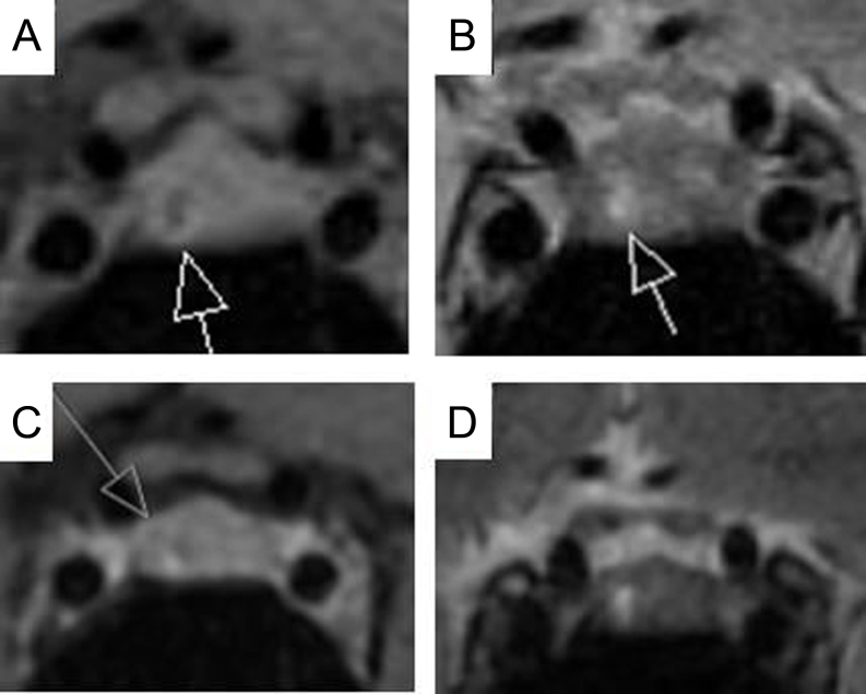
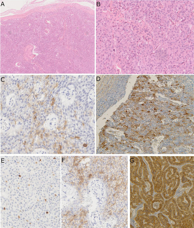
Summary: A male patient with a germline mutation in MEN1 presented at the age of 18 with classical features of gigantism. Previously, he had undergone resection of an insulin-secreting pancreatic neuroendocrine tumour (pNET) at the age of 10 years and had subtotal parathyroidectomy due to primary hyperparathyroidism at the age of 15 years. He was found to have significantly elevated serum IGF-1, GH, GHRH and calcitonin levels. Pituitary MRI showed an overall bulky gland with a 3 mm hypoechoic area. Abdominal MRI showed a 27 mm mass in the head of the pancreas and a 6 mm lesion in the tail. Lanreotide-Autogel 120 mg/month reduced GHRH by 45% and IGF-1 by 20%. Following pancreaticoduodenectomy, four NETs were identified with positive GHRH and calcitonin staining and Ki-67 index of 2% in the largest lesion. The pancreas tail lesion was not removed. Post-operatively, GHRH and calcitonin levels were undetectable, IGF-1 levels normalised and GH suppressed normally on glucose challenge. Post-operative fasting glucose and HbA1c levels have remained normal at the last check-up. While adolescent-onset cases of GHRH-secreting pNETs have been described, to the best of our knowledge, this is the first reported case of ectopic GHRH in a paediatric setting leading to gigantism in a patient with MEN1. Our case highlights the importance of distinguishing between pituitary and ectopic causes of gigantism, especially in the setting of MEN1, where paediatric somatotroph adenomas causing gigantism are extremely rare.
Learning points: It is important to diagnose gigantism and its underlying cause (pituitary vs ectopic) early in order to prevent further growth and avoid unnecessary pituitary surgery. The most common primary tumour sites in ectopic acromegaly include the lung (53%) and the pancreas (34%) (1): 76% of patients with a pNET secreting GHRH showed a MEN1 mutation (1). Plasma GHRH testing is readily available in international laboratories and can be a useful diagnostic tool in distinguishing between pituitary acromegaly mediated by GH and ectopic acromegaly mediated by GHRH. Positive GHRH immunostaining in the NET tissue confirms the diagnosis. Distinguishing between pituitary (somatotroph) hyperplasia secondary to ectopic GHRH and pituitary adenoma is difficult and requires specialist neuroradiology input and consideration, especially in the MEN1 setting. It is important to note that the vast majority of GHRH-secreting tumours (lung, pancreas, phaeochromocytoma) are expected to be visible on cross-sectional imaging (median diameter 55 mm) (1). Therefore, we suggest that a chest X-ray and an abdominal ultrasound checking the adrenal glands and the pancreas should be included in the routine work-up of newly diagnosed acromegaly patients.
Figures
References
Grants and funding
LinkOut - more resources
Full Text Sources
Miscellaneous
