[Value of Combined Detection of Cytokines and Tumor Markers in the Differential Diagnosis of Benign and Malignant Solitary Pulmonary Nodules]
- PMID: 34157802
- PMCID: PMC8246392
- DOI: 10.3779/j.issn.1009-3419.2021.102.20
[Value of Combined Detection of Cytokines and Tumor Markers in the Differential Diagnosis of Benign and Malignant Solitary Pulmonary Nodules]
Abstract
Background: Solitary pulmonary nodule has received increasing attention in recent years. A couple of lung nodules have been recognized as primary malignant tumors, which leads to an urgent need in enhancing the diagnosis of benign/malignant lung nodules at clinical settings. This study aims to explore the value of the combined detection of cytokines and tumor markers in differencing benign and malignant solitary pulmonary nodules in diagnose.
Methods: With 81 solitary pulmonary nodules cases with a clear diagnosis, the general clinical data, nodule imaging features, pathological diagnosis data, serological index cytokine series and tumor marker expression levels were collected in groups. Both single factor and multi-factors analysis were conducted to screen out the serum influence indexes that can predict the malignant probability of lung nodules, and mean while binary logistic regression analysis was used to construct joint indexes; After receiver operating characteristic curve (ROC) was drawn, the area under the curve and the corresponding sensitivity, specificity and positive of each index predicted value, negative predicted value and accuracy could be calculated with a view to determine the statistical significance of area under the curve (AUC).
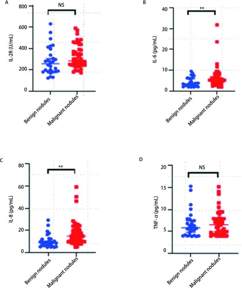
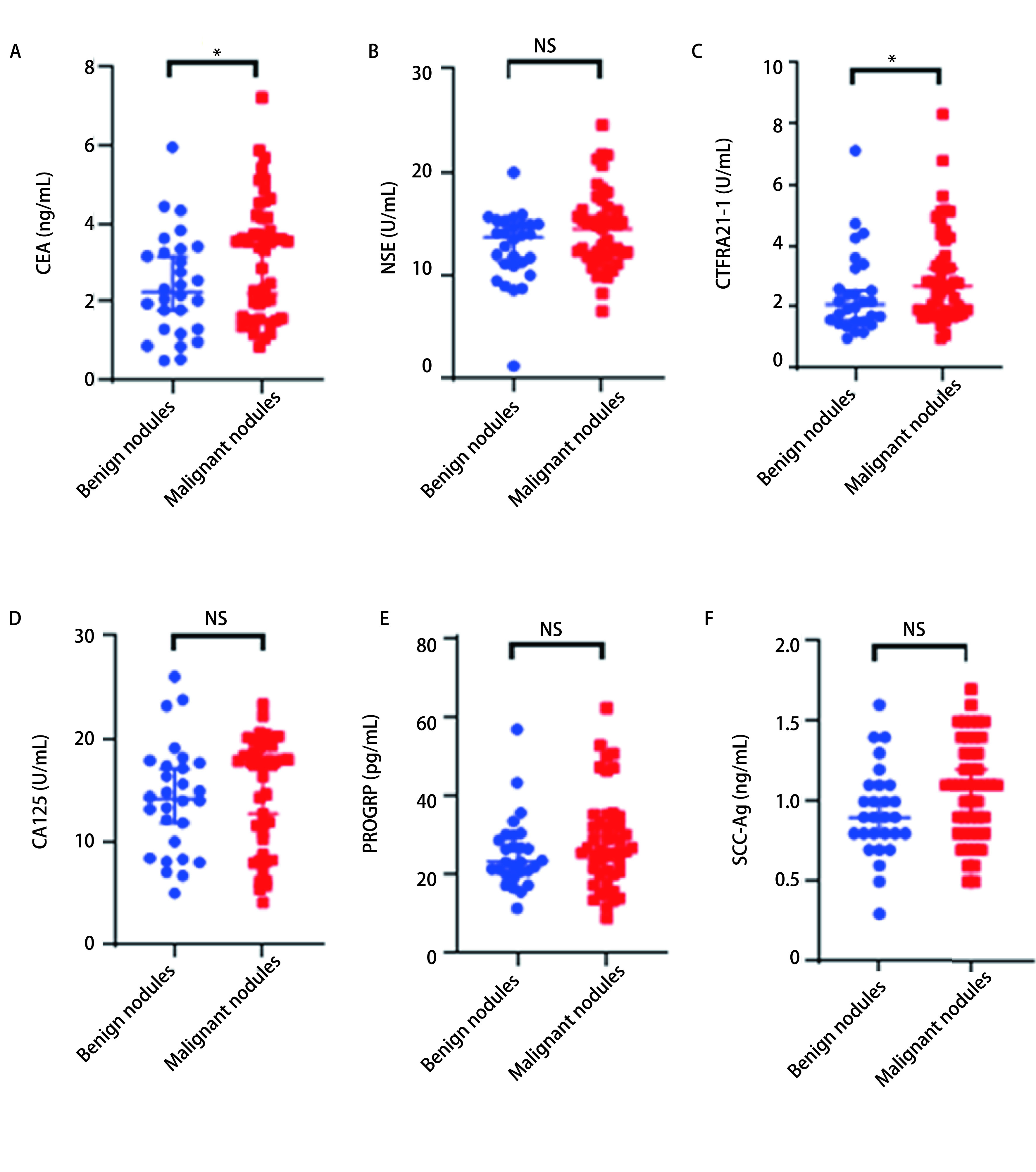
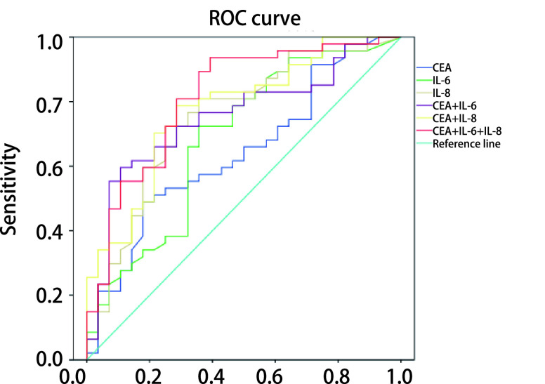
Results: There are differences in the distribution of malignant solitary pulmonary nodules at different locations, with the highest proportion of the right upper lobe (40.4%). The serum levels of carcinoembryonic antigen (CEA), cytokeratin 19 fragment 21-1 (CYFRA21-1), interleukin-6 (IL-6), interleukin-8 (IL-8) in the malignant nodule group were higher than those in the benign nodule group. Logistic regression analysis suggests that CEA, IL-6 and IL-8 are independent risk factors for predicting malignant nodules. ROC curve analysis shows that the areas under the curve of the individual indicators CEA, IL-6 and IL-8 are 0.642, 0.684 and 0.749. The comparison result of the test efficiency of the area under the curve suggests that CEA+IL-6+IL-8 has a larger area under the curve and higher detection efficiency.
Conclusions: CEA, IL-6 and IL-8 are independent risk factors for malignant solitary pulmonary nodules. The combined detection of cytokines and tumor markers has played a role in the differential diagnosis of benign and malignant lung nodules. The diagnostic value of the combined detection of CEA+IL-6+IL-8 is the highest.
【中文题目:细胞因子与肿瘤标志物联合检测对孤立性 肺结节良恶性鉴别诊断的价值】 【中文摘要:背景与目的 近年来,孤立性肺结节(solitary pulmonary nodule, SPN)受到越来越多的关注,部分肺结节被认为是早期肺癌,但如何鉴别肺结节良恶性却是亟待解决的临床难题。本研究旨在探讨细胞因子与肿瘤标志物联合检测对SPN良恶性的鉴别诊断价值,从而提高SPN诊断的准确性。方法 纳入81例诊断明确的SPN患者作为研究对象,收集病例的一般临床资料、结节影像学特征、病理学诊断资料、细胞因子系列和肿瘤标志物表达水平。利用单因素和多因素分析筛选可预测肺结节性质的影响指标,并用二元Logistic回归分析构造联合指标;绘制受试者工作特征曲线(receiver operating characteristic curve, ROC),计算曲线下面积及相应的灵敏度、特异度、阳性预测值、阴性预测值和准确率。结果 一般临床资料分析示恶性结节出现在右肺上叶的比例最高(40.4%)。 恶性结节组中的 癌胚抗原(carcinoembryonic antigen, CEA)、细胞角蛋白19片段(cytokeratin 19 fragment 21-1, CYFRA21-1)、白介素6(interleukin-6, IL-6)和白介素8 (interleukin-8 , IL-8)血清水平高于良性结节组。Logistic回归分析提示,CEA、IL-6、IL-8为预测恶性结节的独立危险因子。ROC曲线分析表明,单项指标CEA、IL-6和IL-8的曲线下面积分别为0.642、0.684和0.749,CEA+IL-6+IL-8联合检测曲线下面积更大,检测效能更高。结论 CEA、IL-6和IL-8为恶性结节的独立危险因素。细胞因子和肿瘤标志物联合检测在SPN良恶性鉴别诊断中具有一定的价值。其中CEA+IL-6+IL-8联合检测的诊断价值最高。 】 【中文关键词:孤立性肺结节;肿瘤标志物;细胞因子;联合检测;诊断】.
Keywords: Combined detection; Cytokines; Diagnosis; Solitary pulmonary nodule; Tumor markers.
Conflict of interest statement
【
Figures
Similar articles
-
Combining serum miRNAs, CEA, and CYFRA21-1 with imaging and clinical features to distinguish benign and malignant pulmonary nodules: a pilot study : Xianfeng Li et al.: Combining biomarker, imaging, and clinical features to distinguish pulmonary nodules.World J Surg Oncol. 2017 May 25;15(1):107. doi: 10.1186/s12957-017-1171-y. World J Surg Oncol. 2017. PMID: 28545454 Free PMC article.
-
[Diagnostic value of serum tumor markers in differentiating malignant from benign solitary pulmonary nodules].Beijing Da Xue Xue Bao Yi Xue Ban. 2014 Oct 18;46(5):707-10. Beijing Da Xue Xue Bao Yi Xue Ban. 2014. PMID: 25331391 Chinese.
-
Diagnostic value of conventional tumor markers in young patients with pulmonary nodules.J Clin Lab Anal. 2021 Sep;35(9):e23912. doi: 10.1002/jcla.23912. Epub 2021 Jul 23. J Clin Lab Anal. 2021. PMID: 34296781 Free PMC article.
-
[Diagnostic approach to solitary pulmonary nodule].Tuberk Toraks. 2005;53(3):307-18. Tuberk Toraks. 2005. PMID: 16258894 Review. Turkish.
-
Application of liquid biopsy in differentiating lung cancer from benign pulmonary nodules (Review).Int J Mol Med. 2025 Jul;56(1):106. doi: 10.3892/ijmm.2025.5547. Epub 2025 May 9. Int J Mol Med. 2025. PMID: 40341969 Free PMC article. Review.
Cited by
-
Application of CT Imaging in Differential Diagnosis and Nursing of Endocrine Tumors.Contrast Media Mol Imaging. 2022 Aug 8;2022:4071081. doi: 10.1155/2022/4071081. eCollection 2022. Contrast Media Mol Imaging. 2022. Retraction in: Contrast Media Mol Imaging. 2023 Oct 4;2023:9831473. doi: 10.1155/2023/9831473. PMID: 36043145 Free PMC article. Retracted.
-
Immunocyte count combined with CT features for distinguishing pulmonary tuberculoma from malignancy among non-calcified solitary pulmonary solid nodules.J Thorac Dis. 2023 Feb 28;15(2):386-398. doi: 10.21037/jtd-22-1024. Epub 2023 Jan 31. J Thorac Dis. 2023. PMID: 36910060 Free PMC article.
-
Diagnostic value of artificial intelligence based on computed tomography (CT) density in benign and malignant pulmonary nodules: a retrospective investigation.PeerJ. 2024 Jan 2;12:e16577. doi: 10.7717/peerj.16577. eCollection 2024. PeerJ. 2024. PMID: 38188164 Free PMC article.
References
-
- Sun KX, Zheng RS, Zhang SW, et al. Analysis of incidence and mortality of malignant tumors in different regions of China in 2015. Zhongguo Zhong Liu. 2019;28(1):1–11.
- 孙 可欣, 郑 荣寿, 张 思维, et al. 2015年中国分地区恶性肿瘤发病和死亡分析. 中国肿瘤. 2019;28(1):1–11. doi: 10.11735/j.issn.1004-0242.2019.01.A001. - DOI
MeSH terms
Substances
LinkOut - more resources
Full Text Sources
Medical