. 2021 Jun 22;11(1):113.
doi: 10.1186/s13578-021-00627-z.

Protein arginine methyltransferase 1 regulates cell proliferation and differentiation in adult mouse adult intestine

Affiliations

Protein arginine methyltransferase 1 regulates cell proliferation and differentiation in adult mouse adult intestine

Lu Xue et al. Cell Biosci. .

Abstract

Background: Adult stem cells play an essential role in adult organ physiology and tissue repair and regeneration. While much has been learnt about the property and function of various adult stem cells, the mechanisms of their development remain poorly understood in mammals. Earlier studies suggest that the formation of adult mouse intestinal stem cells takes place during the first few weeks after birth, the postembryonic period when plasma thyroid hormone (T3) levels are high. Furthermore, deficiency in T3 signaling leads to defects in adult mouse intestine, including reduced cell proliferation in the intestinal crypts, where stem cells reside. Our earlier studies have shown that protein arginine methyltransferase 1 (PRMT1), a T3 receptor coactivator, is highly expressed during intestinal maturation in mouse.

Methods: We have analyzed the expression of PRMT1 by immunohistochemistry and studied the effect of tissue-specific knockout of PRMT1 in the intestinal epithelium.

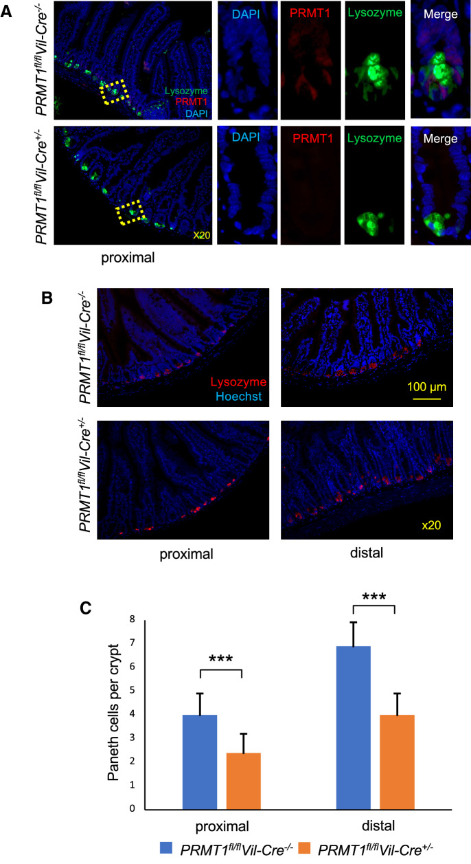
Results: We show that PRMT1 is expressed highly in the proliferating transit amplifying cells and crypt base stem cells. By using a conditional knockout mouse line, we have demonstrated that the expression of PRMT1 in the intestinal epithelium is critical for the development of the adult mouse intestine. Specific removal of PRMT1 in the intestinal epithelium results in, surprisingly, more elongated adult intestinal crypts with increased cell proliferation. In addition, epithelial cell migration along the crypt-villus axis and cell death on the villus are also increased. Furthermore, there are increased Goblet cells and reduced Paneth cells in the crypt while the number of crypt base stem cells remains unchanged.

Conclusions: Our finding that PRMT1 knockout increases cell proliferation is surprising considering the role of PRMT1 in T3-signaling and the importance of T3 for intestinal development, and suggests that PRMT1 likely regulates pathways in addition to T3-signaling to affect intestinal development and/or homeostasis, thus affecting cell proliferating and epithelial turn over in the adult.

Keywords: Adult organ-specific stem cell; Histone arginine methyltransferase; Intestine; Thyroid hormone receptor; Transcription coactivator.


Conflict of interest statement

The authors declare no competing interests.

Figures

Fig. 1
Fig. 1
Generation and confirmation of intestinal epithelium-specific PRMT1 conditional knockout mice. A Schematic diagram of the wild-type PRMT1 allele with LoxP sites (white arrowheads) flanking exons 5 and 6, and an FRT-flanked β-galactosidase/neomycin (β-gal/neo) cassette, which can be removed by crossing the PRMT1LoxP mice with Flp (Flipase)-expressing mice to produce mice containing PRMT1fl allele. The resulting PRMT1fl-containing mice can be crossed with mice expressing the Cre recombinase under the control of the Villin promoter to remove the floxed exons 5 and 6, thus knocking out this PRMT1 allele. B PCR-genotyping of genomic DNA extracted from tail biopsies. Homozygous PRMT1fl/fl mice were crossed with mice that were heterozygous in Villin Cre and floxed PRMT1 (PRMT1fl/+Cre+/−). The resulting animals as well as wild type (WT) were genotyped by PCR with a PRMT1 primer combination that distinguishes WT, PRMT1fl/fl and PRMT1fl/+ genotypes, which produced bands in agarose gel of 377 bp for PRMT1 floxed allele and 277 bp for WT PRMT1 allele., respectively. The presence or absence of Cre was also analyzed by PCR with a specific primer pair to produce a band of about 1000 bp
Fig. 2
Fig. 2
Intestinal epithelium-specific deletion of PRMT1 alters the structural characteristics of adult intestine. A PRMT1 knockout does not affect absolute intestinal length. The length of the intestine was measured for 8-week-old adult wild type (PRMT1fl/flVil-Cre−/−) or intestinal epithelium-specific PRMT1 knockout (PRMT1fl/flVil-Cre+/−) mice. B PRMT1 knockout increases the relative length of the intestine normalized by body weight. The length of the intestine (cm) as in A was normalized against the body weight of the animals (grams). CE PRMT1 knockout increases the crypt length. The intestine from 8-week-old wild type or PRMT1 knockout mice were dissected and stained with H&E staining (C) and the length of the villus (D) or crypt (E) were measured for both distal and proximal small intestine from multiple sections per animal. Note that the crypt length was increased in both distal and proximal small intestine. The villus length was not altered in the proximal small intestine and slightly decreased in the distal small intestine. For both PRMT1 knockout and control littermates, n = 4. *p < 0.05, **p < 0.01, ***p < 0.001. ns no significant
Fig. 3
Fig. 3
PRMT1 knockout does not affect intestinal stem cell number. The proximal small intestine of 13-week-old adult wild type (PRMT1fl/flVil-Cre−/−) or intestinal epithelium-specific PRMT1 knockout (PRMT1fl/flVil-Cre+/−) mice were sectioned and analyzed by in situ hybridization with Olfm4 probe for crypt base stem cells (A). The number of stem cells were counted visually from multiple sections per animal (B). For both PRMT1 knockout and wild type littermates, n = 4. *p < 0.05, **p < 0.01
Fig. 4
Fig. 4
Intestinal epithelium-specific deletion of PRMT1 increases cell proliferation in the crypt. A High levels of PRMT1 are present in the crypt base and transit amplifying cells in wild type small intestine. 8-week-old adult wild type (PRMT1fl/flVil-Cre−/−) or PRMT1 knockout (PRMT1fl/flVil-Cre+/−) mice were injected with EdU to label proliferating cells. The proximal small intestine was then isolated for detection of EdU and PRMT1 expression by immunohistochemistry. The DNA was stained with Hoechst. Note that EdU labeled mostly transit amplifying cells which had high levels of PRMT1 in the wild type animals. There were also PRMT1-expressing cells at the bottom of the crypt where stem cells reside. Expectedly, no PRMT1 proteins were found in the epithelium of PRMT1 knockout mice (PRMT1fl/flVil-Cre+/−). Note the increased cell proliferation in the crypts of PRMT1 knockout mice. B, C PRMT1 knockout increases cell proliferation in both proximal and distal small intestine. Both proximal and distal small intestine of wild type and PRMT1 knockout mice were analyzed by EdU labeling and Hoechst staining (B) and the number of proliferating transit amplifying cells were quantified from multiple sections per animal (C). For both PRMT1 knockout and control wild type littermates, n = 4. ***p < 0.001
Fig. 5
Fig. 5
Intestinal epithelium-specific deletion of PRMT1 increases Goblet cells in the crypt. The proximal and distal small intestine of 8-week-old adult wild type (PRMT1fl/flVil-Cre−/−) or PRMT1 knockout (PRMT1fl/flVil-Cre+/−) mice were sectioned and stained with Alcian Blue to detect the goblet cells (A). The number of Goblet cells in the villi and crypts were counted from multiple sections per animal and graphed (B, C). Note that Goblet cell number was increased in the crypt but not villus of both distal and proximal intestine in PRMT1 knockout mice. For both PRMT1 knockout and control wild type littermates, n = 4. ***p < 0.001. ns no significant
Fig. 6
Fig. 6
Intestinal epithelium-specific deletion of PRMT1 reduces Paneth cells in the crypt. A PRMT1 protein is low or absent in Paneth cells in the adult wild type intestine. The small intestine of 8-week-old adult wild type (PRMT1fl/flVil-Cre−/−) or PRMT1 knockout (PRMT1fl/flVil-Cre+/−) mice were sectioned and stained with anti-PRMT1 antibody for PRMT1 expression (red) or anti-lysozyme antibody for Paneth cells in the crypt (green). Note that the signals for PRMT1 and lysozyme were in different cells in the wild type intestine, with PRMT1-expressing cells surrounding the lysozyme-positive Paneth cells, which is consistent with PRMT1-expressing cells being the crypt base stem cells). Expectedly, PRMT1 was not detected in the knockout epithelium. B, C PRMT1 knockout reduces Paneth cells in both proximal and distal small intestine. The proximal and distal small intestine of wild type and PRMT1 knockout mice were stained with anti-lysozyme antibody for Paneth cells and Hoechst for DNA (B) and the number of Paneth cells were quantified from multiple sections per animal (C). For both PRMT1 knockout and control wild type littermates, n = 4. ***p < 0.001. ns no significant
Fig. 7
Fig. 7
PRMT1 knockout increases cell death in the intestine. The proximal and distal small intestine of 8-week-old adult wild type (PRMT1fl/flVil-Cre−/−) or intestinal epithelium-specific PRMT1 knockout (PRMT1fl/flVil-Cre+/−) mice were sectioned and analyzed with TUNEL for apoptotic cells (red) and DAPI for DNA (blue) (A). The apoptotic cells per villus-crypt unit were quantified from multiple sections per animal (B). For both PRMT1 knockout and control wild type littermates, n = 4. *p < 0.05, **p < 0.01
Fig. 8
Fig. 8
Intestinal epithelium-specific deletion of PRMT1 accelerates the epithelial migration along the crypt-vilus axis. The proximal (A) or distal (B) small intestine of 8-week-old wild type (PRMT1fl/flVil-Cre−/−) or PRMT1 knockout (PRMT1fl/flVil-Cre+/−) mice were sectioned at indicated times after EdU injection. Note that in both proximal and distal intestine, essentially all EdU-labeled cells were located in the crypts at 2 h after EdU injection in both wild type and knockout animals. 24 h after EdU injection, EdU-labeled cells migrated up to the lower middle of villus, with the faster migration in the knockout mice. 48 h after EdU injection, EdU-labeled cells migrated to the middle (for proximal intestine) or near the top (for distal intestine) of villus in wild type animals but in the knockout animals, most of the labeled cells were lost, suggesting that they had migrated to the very top of the villus and undergone apoptosis

Similar articles

Cited by

References

    1. MacDonald WC, Trier JS, Everett NB. Cell proliferation and migration in the stomach, duodenum, and rectum of man: radioautographic studies. Gastroenterology. 1964;46:405–417. doi: 10.1016/S0016-5085(64)80102-5. - DOI - PubMed
    1. Toner PG, Carr KE, Wyburn GM. The digestive system: an ultrastructural atlas and review. London: Butterworth; 1971.
    1. van der Flier LG, Clevers H. Stem cells, self-renewal, and differentiation in the intestinal epithelium. Annu Rev Physiol. 2009;71:241–260. doi: 10.1146/annurev.physiol.010908.163145. - DOI - PubMed
    1. Sancho E, Eduard Batlle E, Clevers H. Signaling pathways in intestinal development and cancer. Annu Rev Cell DevBiol. 2004;20:695–723. doi: 10.1146/annurev.cellbio.20.010403.092805. - DOI - PubMed
    1. Harper J, Mould A, Andrews RM, Bikoff EK, Robertson EJ. The transcriptional repressor Blimp1/Prdm1 regulates postnatal reprogramming of intestinal enterocytes. Proc Natl Acad Sci USA. 2011;108(26):10585–10590. doi: 10.1073/pnas.1105852108. - DOI - PMC - PubMed

LinkOut - more resources