. 2021 Sep;31(9):998-1010.
doi: 10.1038/s41422-021-00524-7. Epub 2021 Jun 22.

Endogenous reverse transcriptase and RNase H-mediated antiviral mechanism in embryonic stem cells

Affiliations

Endogenous reverse transcriptase and RNase H-mediated antiviral mechanism in embryonic stem cells

Junyu Wu et al. Cell Res. 2021 Sep.

Abstract

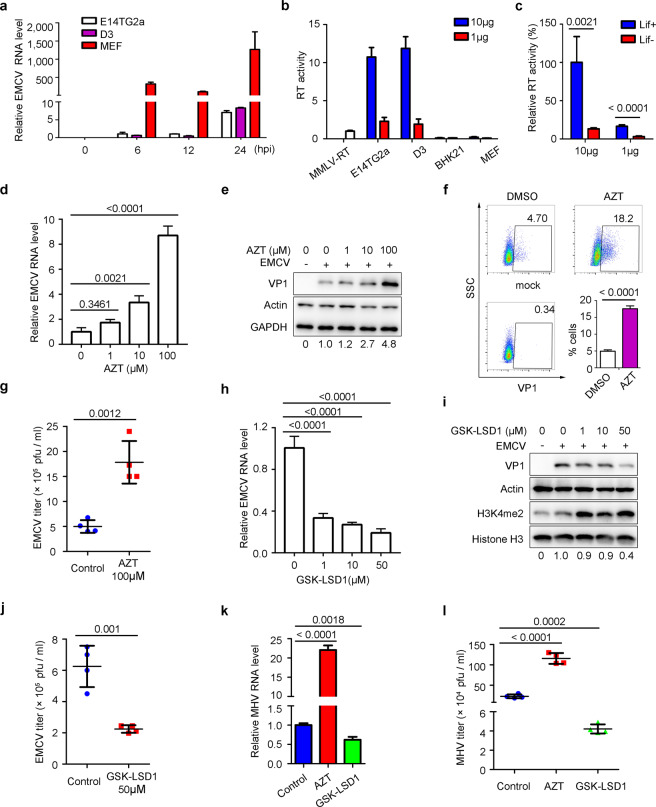
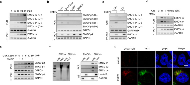
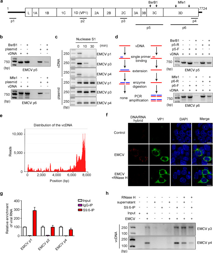
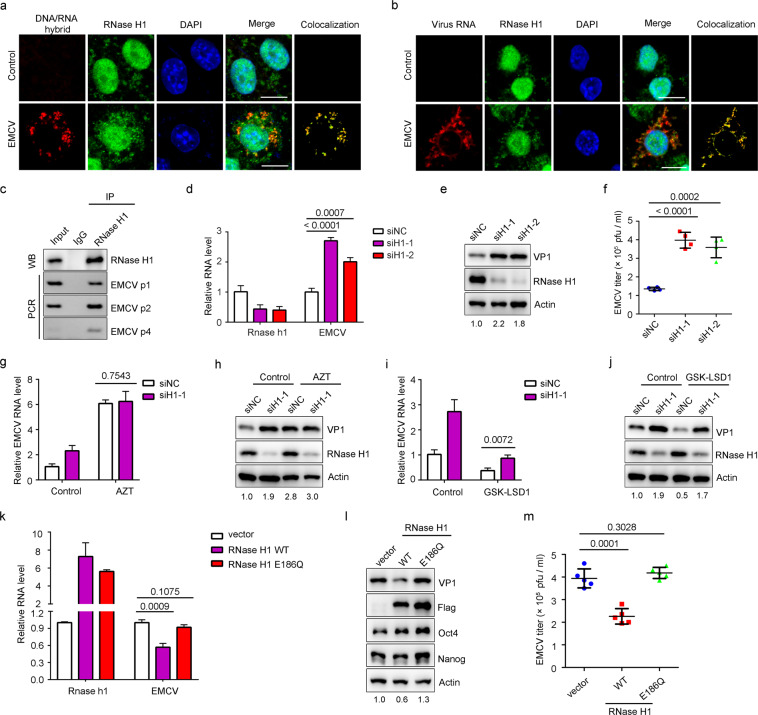
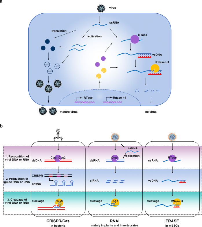
Nucleic acid-based systems play important roles in antiviral defense, including CRISPR/Cas that adopts RNA-guided DNA cleavage to prevent DNA phage infection and RNA interference (RNAi) that employs RNA-guided RNA cleavage to defend against RNA virus infection. Here, we report a novel type of nucleic acid-based antiviral system that exists in mouse embryonic stem cells (mESCs), which suppresses RNA virus infection by DNA-mediated RNA cleavage. We found that the viral RNA of encephalomyocarditis virus can be reverse transcribed into complementary DNA (vcDNA) by the reverse transcriptase (RTase) encoded by endogenous retrovirus-like elements in mESCs. The vcDNA is negative-sense single-stranded and forms DNA/RNA hybrid with viral RNA. The viral RNA in the heteroduplex is subsequently destroyed by cellular RNase H1, leading to robust suppression of viral growth. Furthermore, either inhibition of the RTase activity or depletion of endogenous RNase H1 results in the promotion of virus proliferation. Altogether, our results provide intriguing insights into the antiviral mechanism of mESCs and the antiviral function of endogenized retroviruses and cellular RNase H. Such a natural nucleic acid-based antiviral mechanism in mESCs is referred to as ERASE (endogenous RTase/RNase H-mediated antiviral system), which is an addition to the previously known nucleic acid-based antiviral mechanisms including CRISPR/Cas in bacteria and RNAi in plants and invertebrates.


Conflict of interest statement

The authors declare no competing interests.

Figures

Fig. 1
Fig. 1. Role of endogenous RTase in antiviral responses in mESCs.
a mESCs were resistant to virus infection. mESCs (E14TG2a and D3) and MEFs were infected with EMCV (MOI = 1) for the indicated time periods. The RNA level of EMCV was determined by quantitative real-time PCR (qRT-PCR). b mESCs contain higher endogenous RTase activity than somatic cells. The endogenous RTase activity of D3, E14TG2a, BHK21 and MEF cell extracts was measured as described in Materials and Methods using MS2 RNA as templates. The levels of MS2 cDNA were determined by qRT-PCR with commercial reverse transcriptase, M-MLV, as positive control. The RTase activity was calculated relative to 1 U of M-MLV. c The endogenous RTase activity decreased following the differentiation of mESCs. E14TG2a cells were cultured in the medium with or without Lif for 7 days. Cells were lysed and the RTase activity was measured. The relative RTase activity was presented by setting the Lif + (10 μg) group as 100%. dg Inhibition of endogenous RTase activity by AZT promoted virus infection in mESCs. E14TG2a cells were infected with EMCV (MOI = 1) after treatment with AZT at the indicated concentrations for 6 h. The RNA level of EMCV was determined by qRT-PCR (d) and the protein level of EMCV VP1 was analyzed by immunoblotting (e). Actin and GAPDH were used as loading control. Intensity of VP1 bands was quantitated by ImageJ and normalized to intensity of actin bands and the result is shown at the bottom. The viral infection rates were detected by flow cytometric analysis with VP1 antibody (f). The viral titers in the medium were measured by plaque assay (g). hj GSK-LSD1 inhibited virus infection in mESCs. E14TG2a cells were pre-treated with GSK-LSD1 at the indicated concentrations for 24 h and were infected with EMCV (MOI = 1) for another 24 h. The RNA level of EMCV was determined by qRT-PCR (h). The protein level of EMCV VP1 and the level of histone H3K4me2 were analyzed by immunoblotting (i). Viral titers in the medium were measured by plaque assay (j). k, l The role of endogenous RTase in MHV infection. The E14TG2a cells were pre-treated with 100 μM AZT or 50 μM GSK-LSD1 and infected with MHV (MOI = 1) for 24 h. The RNA level of MHV was determined by qRT-PCR (k) and viral titers were measured by plaque assay (l). Data are representative of three independent experiments (e, i). The graphs represent means ± standard deviation (SD) from three (a, d, f, h) or four (b, c, g, j, k, l) independent replicates measured in triplicate. Statistics were calculated by the two-tailed unpaired Student’s t-test (c, f, g, j, k, l) or one-way ANOVA with Tukey’s post hoc tests (d, h).
Fig. 2
Fig. 2. Generation of viral DNA in the cytoplasm of infected mESCs.
a Kinetics of the vDNA synthesis in mESCs. E14TG2a cells were infected with EMCV, followed by extraction of total DNA and RNA at 0–48 hpi (hours after infection). Total DNA was treated with Turbo DNase (D+ ) or not (D–). The virus RNA and vDNA levels were analyzed by RT-PCR (lower panel) and PCR (upper panel), respectively. GAPDH was used as loading control. The positions of the primers were marked in Fig. 3a. b D3, E14TG2a, MEF and BHK21 cells were infected with EMCV for 24 h. The virus RNA and vDNA levels were analyzed by RT-PCR (lower panel) and PCR (upper panel), respectively. c The level of vDNA decreased following the differentiation of mESCs. mESCs cultured in the medium with or without Lif for 7 days were infected by EMCV for 24 h. The virus RNA and vDNA levels were analyzed, respectively. d AZT inhibits vDNA synthesis in mESCs. E14TG2a cells were infected with EMCV (MOI = 1) after treatment with AZT at the indicated concentrations for 6 h. The virus RNA and vDNA levels were analyzed, respectively. e GSK-LSD1 promotes vDNA production in mESCs. E14TG2a cells were pre-treated with GSK-LSD1 at the indicated concentrations for 24 h and were infected with EMCV for another 24 h. The virus RNA and vDNA levels were analyzed, respectively. f The vDNA is mainly localized in the cytoplasm. The cytoplasmic and nuclear fractions were isolated from E14TG2a cells with or without virus infection. The DNA of each fraction was extracted and analyzed by agarose gel electrophoresis (left panel). The vDNA was detected by PCR with the indicated primers (right upper panel). Lamin B and GAPDH were used as nuclear and cytoplasmic markers by western blotting, respectively (right lower panel). g E14TG2a cells infected with EMCV (MOI = 1, 24 hpi) were examined for the localization of vDNA (red) and VP1 (green) by FISH. Nuclei were stained with DAPI (blue). Scale bars, 10 μm. Data are representative of three independent experiments (ag).
Fig. 3
Fig. 3. Formation of DNA/RNA heteroduplex by interaction of single-stranded antisense vDNA (vcDNA) with viral RNA.
a Schematics of EMCV viral genome. The bottom indicates the positions of the PCR primer pairs used for the detection of vDNA. The arrows above the genome show the sites of restriction endonucleases, BsrBI and MfeI. b The vDNA is resistant to DNA restrictive endonucleases. The vDNA was extracted from E14TG2a cells infected with EMCV and incubated with restrictive endonuclease BsrBI or MfeI for 1 h. The integrity of DNA was evaluated by PCR with the indicated primers. The plasmid pCMV-rNJ08, which contains full-length EMCV genomic sequence, was used as positive control of dsDNA. c The vDNA is single-stranded. The vDNA and plasmid pCMV-rNJ08 were incubated with nuclease S1 at 1 U for the indicated time periods. The integrity of DNA was evaluated by PCR with the indicated primers. d The vDNA is complementary with the virus genomic sequence (vcDNA). Unidirectional primer extension was performed as described in Materials and Methods with the indicated primers. The products were then digested by the dsDNA restrictive endonucleases and PCR was performed to evaluate the integrity. e The distribution of vcDNA. The vcDNA was enriched with RNA probes targeting the complementary sequence of the virus genome and was sequenced as described in Materials and Methods, n = 2. f DNA/RNA hybrids were accumulated in virus-infected mESCs. E14TG2a cells were infected with EMCV (MOI = 1, 24 hpi). Cells were mock-treated or pre-treated with RNase H for 1 h at room temperature. The DNA/RNA hybrids (S9.6 antibody, red) and VP1 (green) were visualized by immunofluorescent staining. Nuclei were stained with DAPI (blue). Scale bars, 10 μm. g, h The vcDNA forms DNA/RNA hybrids with viral RNA. The cytoplasmic nucleic acids were extracted from E14TG2a cells infected with EMCV (MOI = 1) for 48 h. The DRIP experiments were performed as described in Materials and Methods with S9.6 antibody. Normal mouse IgG was used as negative control. The viral RNA was analyzed by qRT-PCR (g) and the vcDNA level in the supernatant or immunoprecipitates was detected by PCR (h). Data are representative of three independent experiments (bd, f, h). In g, the graph represents means ± SD from three independent replicates measured in triplicate.
Fig. 4
Fig. 4. Engagement of RNase H1 cleavage in antiviral activity of vcDNA in mESCs.
a DNA/RNA hybrids induced by virus infection recruit endogenous RNase H1. The infected and uninfected E14TG2a cells were fixed and stained with RNA/DNA hybrid-specific S9.6 antibody (red) and RNase H1 antibody (green). Nuclei were stained with DAPI (blue). Scale bars, 10 μm. The colocalization area was shown in the right panel. b Viral RNA is colocalized with RNase H1. Viral RNA was visualized by RNAscope (red), followed by immunostaining with RNase H1 (green) and DAPI (blue). Scale bars, 10 μm. The colocalization area was shown in the right panel. c RNase H1 binds to vcDNA. EMCV-infected E14TG2a cell lysates were immunoprecipitated with anti-RNase H1 antibody. The pull-down efficiency was validated by western blotting (upper panel). The enrichment of vcDNA was analyzed by PCR with the indicated primers (lower panel). Normal rabbit IgG was used as negative control. df Depletion of RNase H1 promoted virus infection. E14TG2a cells were infected with EMCV (MOI = 1) after transfection with siRNAs for 36 h. The RNA level of EMCV was determined by qRT-PCR (d), the protein level of VP1 was analyzed by immunoblotting (e), and viral titers were measured by plaque assay (f). Intensity of VP1 bands was quantitated by ImageJ and normalized to intensity of actin bands. The result is shown at the bottom. g, h RNase H1 functions downstream of endogenous RTase to inhibit viral infection. The siRNA-transfected E14TG2a cells were treated with 100 μM AZT for 6 h and then infected with EMCV for another 24 h. Both the levels of viral RNA (g) and the VP1 protein (h) were analyzed. i, j The antiviral function of GSK-LSD1 is partially dependent on RNase H1. The siRNA-transfected E14TG2a cells were pre-treated with 50 μM GSK-LSD1 for 24 h before infection with EMCV. Both the levels of viral RNA (i) and the VP1 protein (j) were analyzed. km RNase H1 inhibition of virus infection relys on its enzyme activity. The wild type and an enzymatically inactive mutant (E186Q) of mouse RNase H1 were transfected into E14TG2a cells followed by virus infection. The levels of viral RNA (k), the VP1 protein (l) and the viral titers (m) were analyzed. Data are representative of three independent experiments (ac, e, h, j, l). The graphs represent means ± SD from three (d, g, i, k), four (f) or five (m) independent replicates measured in triplicate. Statistics were calculated by the two-tailed unpaired Student’s t-test.
Fig. 5
Fig. 5. The proposed mechanistic model of ERASE and its comparison with RNAi and CRISPR/Cas antiviral mechanisms.
a Working model of ERASE in mESCs. When facing RNA virus infection, the endogenous RTase catalyzes the production of vcDNA from the viral RNA. The vcDNA forms DNA/RNA hybrid structures with viral RNA. Then, the hybrids recruit endogenous RNase H1 to hydrolyze the viral RNA and inhibit virus replication. b The nucleic acid-based antiviral defense generally consists of three important steps: the recognition of viral nucleic acids, the generation of guide RNA or DNA, and the cleavage of viral nucleic acids. CRISPR/Cas is RNA-guided DNA cleavage by Cas enzymes to prevent DNA virus infection in bacteria. RNAi is RNA-guided RNA cleavage by Ago to defend against RNA virus infection mainly in plants and invertebrates. ERASE, in this report, is DNA-mediated RNA cleavage by RNase H to restrict RNA virus infection in mESCs. The ERASE activity in other mammalian species awaits future investigation.

Comment in

References

    1. Horvath P, Barrangou R. CRISPR/Cas, the immune system of bacteria and archaea. Science. 2010;327:167–170. doi: 10.1126/science.1179555. - DOI - PubMed
    1. Guo Z, Li Y, Ding SW. Small RNA-based antimicrobial immunity. Nat. Rev. Immunol. 2019;19:31–44. doi: 10.1038/s41577-018-0071-x. - DOI - PubMed
    1. Yan N, Chen ZJ. Intrinsic antiviral immunity. Nat. Immunol. 2012;13:214–222. doi: 10.1038/ni.2229. - DOI - PMC - PubMed
    1. tenOever BR. The evolution of antiviral defense systems. Cell Host Microbe. 2016;19:142–149. doi: 10.1016/j.chom.2016.01.006. - DOI - PubMed
    1. Burke DC, Graham CF, Lehman JM. Appearance of interferon inducibility and sensitivity during differentiation of murine teratocarcinoma cells in vitro. Cell. 1978;13:243–248. doi: 10.1016/0092-8674(78)90193-9. - DOI - PubMed

Publication types