. 2021 May 31:9:25151355211017119.
doi: 10.1177/25151355211017119. eCollection 2021.

Generating CAR T cells from tumor-infiltrating lymphocytes

Affiliations

Generating CAR T cells from tumor-infiltrating lymphocytes

Jane K Mills et al. Ther Adv Vaccines Immunother. .

Abstract

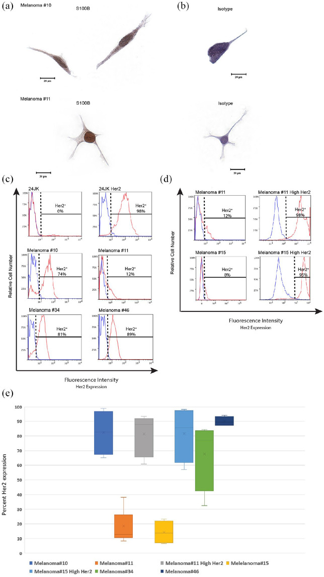
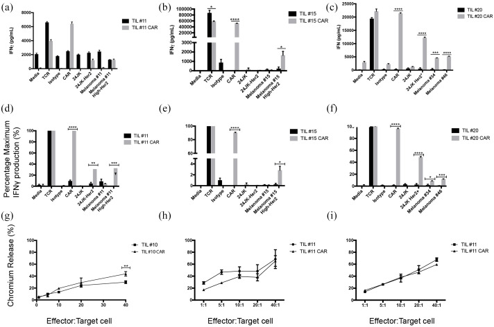
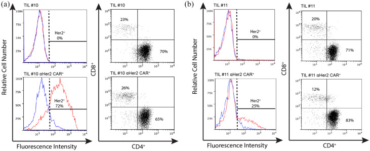
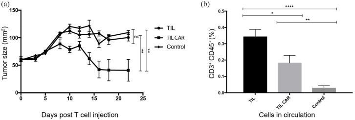
Background: Tumor-infiltrating lymphocytes (TILs) and chimeric antigen receptor (CAR) T-cell therapies have demonstrated promising, though limited, efficacy against melanoma. Methods: We designed a model system to explore the efficacy of dual specific T cells derived from melanoma patient TILs by transduction with a Her2-specific CAR. Results: Metastatic melanoma cells in our biobank constitutively expressed Her2 antigen. CAR-TIL produced greater amounts of IFN compared with parental TIL, when co-cultured with Her2 expressing tumor lines, including autologous melanoma tumor lines, although no consistent increase in cytotoxicity by TIL was afforded by expression of a CAR. Results of an in vivo study in NSG mice demonstrated tumor shrinkage when CAR-TILs were used in an adoptive cell therapy protocol. Conclusion: Potential limitations of transduced TIL in our study included limited proliferative potential and a terminally differentiated phenotype, which would need addressing in further work before consideration of clinical translation.

Keywords: Her2; autologous tumor; chimeric antigen receptor; immunotherapy; melanoma.


Conflict of interest statement

Conflict of interest statement: The authors declare that there is no conflict of interest.

Figures

Figure 1.
Figure 1.
TILs are phenotypically diverse. TILs were thawed from the biobank and activated using CD3/28 beads and IL-2 600 IU/ml in CM. IL-2 was replaced at 2–3 day intervals. Each flow cytometry plot demonstrates the cell phenotype for a different patient, designated by patient #number [(a) TIL#10, (b) TIL#11, (c) TIL#15, (d) TIL#20]. Cell populations were gated on morphology, viability, CD3+ and stained for CD4+ and CD8+. CM, complete media; TIL, tumor-infiltrating lymphocyte.
Figure 2.
Figure 2.
TIL cultures had variable proliferation and terminal differentiation. 1 × 106 T cells were activated with CD3/28 Dynabeads and IL-2 (600 IU/ml). (a) TIL proliferation rates are demonstrated for individual patients designated by patient #number. (b) TIL proliferation rates are compared with allogeneic donor PBMCs (n = 4 PBMC cultures, 6 TIL cultures. p < 0.0001 by one-way analysis of variance). (c) TIL#11, #20 and allogeneic PBMCs were thawed, activated using CD3/28 beads, labeled with CTV and cultured in complete media with IL-2 (600 IU/ml). Cells were then analyzed for CTV fluorescence daily until day 7 for TIL or day 6 for PBMC. TIL#11 and #20 were cultured with IL-2 and IL-7 (10 ng/ml) for comparison. Cell proliferation, as seen from CTV dilution over time, was similar irrespective of whether IL-2 alone or IL-2+IL-7 was used in culture (results of a single experiment). (d) CD4+, and (e) CD8+, demonstrate TIL subtypes for TIL#11 with and without the addition of IL-7 to media. Cells were gated on morphology, viability, CD3+, CD4+ or CD8+. Naïve T cells: CD45RA+ CCR7+; TEMRA: CD45RA+ CCR7; TCM: CD45RA CCR7+; TEM: CD45RA CCR7. Similar proportions of cell subsets were observed in IL-2- and IL-2/IL-7-supplemented cultures (results from a single experiment). CTV, cell trace violet; PBMC, peripheral blood mononuclear cell; TCM, central memory T cell; TEM, effector memory T cell; TEMRA, effector memory T cell expressing CD45RA; TIL, tumor-infiltrating lymphocyte.
Figure 3.
Figure 3.
TILs can be successfully transduced with anti-Her2 CAR. TILs were thawed and activated using CD3/28 beads with IL-2 (600 IU/ml) and transduced with retroviral vector in the presence of retronectin and selected with G418 for expression of anti-He2-CAR. Non-transduced TILs underwent the transduction protocol in the presence of an empty viral vector. Cells were examined for CAR expression using flow cytometry. TIL-CAR were stained with c-Myc tag (red), with an isotype (IgG2A; blue). Cells are gated on morphology, viability, CD3+ and c-Myc tag staining. CAR expression was calculated based on <1% staining of the non-transduced CAR stained with c-Myc tag. (a) Maximal staining of transduction of TIL#10 and (b) TIL#11 transduced with anti-Her2 CAR. (c) CAR expression using flow cytometry shown for multiple transductions. Results are expressed with mean and SEM for each patient. (d) CAR density on transduced T cells using a proprietary bead standard kit (Simply Cellular Beads, BANGSLABS laboratories). CAR density on TILs was pooled and data shown ± SEM performed in triplicate wells. CAR, chimeric antigen receptor; PBMC, peripheral blood mononuclear cell; TIL, tumor-infiltrating lymphocyte
Figure 4.
Figure 4.
Transduced TIL#10 retain CAR expression following REP. TILS were thawed and activated using CD3/28 beads with IL-2 (600 × IU/ml). Five days following activation they were transduced with retroviral vector in the presence of retronectin and selected with G418 for expression of anti-He2-CAR. Non-transduced TILs underwent the transduction protocol in the presence of an empty viral vector to control for any changes induced by the process of transduction. Following confirmation of successful transduction and CAR expression using flow cytometry, to increase cell numbers for experiments, TILs underwent a second activation using REP. REP is a restimulation process utilizing soluble OKT3 and irradiated donor PBMCs. Cells were examined for CAR expression using flow cytometry. Data are shown for c-Myc tag (αHer2 CAR; red) with isotype control (blue). (a) TIL#10 expressed the CAR (92%) following transduction after CD3/28 bead activation and G418 selection. (b) Following further expansion, by REP, cells maintain CAR expression (73%). Control cells transduced with an empty viral vector maintain negative expression for CAR pre-REP (c) and post-REP (d). Cells are gated on cell morphology, viability, CD3+ and stained for c-Myc tag expression (CAR) or isotype (IgG2A; control). CAR, chimeric antigen receptor; OKT3, anti-CD3; PBMC, peripheral blood mononuclear cell; REP, rapid expansion protocol; TIL, tumor-infiltrating lymphocyte.
Figure 5.
Figure 5.
Melanoma tumor cells constitutively express Her2 antigen. (a-b) Melanoma tumor cells #10 and #11 were cultured onto slides and fixed using formaldehyde and stained for S100B. Images were taken using an Olympus BX51 Microscope at ×40 magnification and captured using SpotFire software. Single representative cells are presented stained with either an S100B-specific antibody (a) or isotype control (b). (c) Tumor lines were examined for Her2 antigen by flow cytometry. Cells were gated on morphology, viability and Her2 antigen expression. Cells that express Her2 antigen (red) are demonstrated against an isotype for Her2 (blue). 24JK tumor lines negative and positive for Her2 expression were used as additional controls. (d) Cell lines of melanoma tumors #11 and #15 were transduced using lipofectamine transduction protocols and sorted for Her2 expression. Control lines with 24JK parental and Her2 expressing tumor lines are shown. Melanoma tumor cell lines from patients #11 and #15 are shown before transduction (Melanoma#) and after transduction and sorting (Melanoma# High Her2). Her2 expressing cells are demonstrated in red and isotype staining is demonstrated in blue. (e) Box plot demonstrating range of Her2 antigen expression for each patient cell line (n = ⩾4 observations per patient).
Figure 6.
Figure 6.
TILs transduced with anti-Her2 CAR respond against autologous melanoma tumor cell lines. (a)–(f) IFNγ production of TILs transduced with anti-Her2 CAR compared with non-transduced TILs. (a)–(c) Raw data values. (a) TIL#11 co-cultured with a panel of targets, including both parental (Melanoma#11) and high expressing Her2 tumor lines (Melanoma#11 High Her2); (b) TIL#15 co-cultured with a panel of targets, including parental (Melanoma#15) and high expressing Her2 tumor lines (Melanoma#15 High-Her2); (c) TIL#20 co-cultured with a panel of targets, including HLA matched melanoma tumor lines. Data are presented as mean ± SEM performed in triplicate. (d)–(f) Normalized values of data from panels (a)–(c); we defined T-cell responses to CD3 ligation as “maximal” (100%) and presented responses to other stimuli as a proportion of maximum. (g)–(i) Cytotoxic activity measured using 51Cr release assay. (g) TIL#10 transduced with anti-Her2 CAR were compared with TILs transduced with an empty viral vector control against autologous tumor cell targets. (h) TIL#11 (TIL-CAR versus TIL) compared against parental tumor cell line (mean Her2 antigen expression 11%) and (i) TIL#11 against autologous tumor lines transduced for high expression of Her2 antigen (mean Her2 antigen expression 98%). Statistical significance was determined using unpaired Student’s t-test of triplicate wells from a single experiment (*p ⩽ 0.05, **p ⩽ 0.01, ***p ⩽ 0.001, ****p ⩽ 0.0001). CAR, chimeric antigen receptor; TIL, tumor-infiltrating lymphocyte; TCR, transgenic T-cell receptor
Figure 7.
Figure 7.
Comparing cell lines for transduction efficiency and phenotype. Transduction efficiency comparing TILs transduced with an empty vector (control) and with anti-Her2 CAR (Her2 CAR) with corresponding cell phenotype. (a) CAR expression for TIL#10 transduced with the empty vector (control) and Her2 CAR (shown in red) and isotype (shown in blue). The phenotypes TIL#10 are shown. (b) CAR expression for TIL#11 transduced with empty vector (control) and Her2 CAR. Cell phenotype of TIL#11 is shown. Cells are gated on morphology, viability, CD3+, CD4+ and CD8+. CAR, chimeric antigen receptor; TIL, tumor-infiltrating lymphocyte.
Figure 8.
Figure 8.
ACT using TIL transduced with anti-Her2 CAR inhibited tumor growth in mice. (a) ACT using 1 × 107 TILs transduced with anti-Her2 CAR or non-transduced TILs was delivered into NSG mice bearing established (>50 mm2) xenografts of an autologous patient melanoma tumor cell line. A control (untreated group) was included for comparison. Time is measured from the day ACT was performed (day 0). Treated mice also received five intraperitoneal injections of IL-2 (50,000 IU/injection per mouse) over 3 days. Tumor growth was significantly inhibited by ACT using CAR-transduced T cells. Results are from a single experiment, with seven mice per treatment group and nine mice for control group. Statistical significance was determined using Student’s t-test. (b) A greater proportion of non-transduced TILs was detected in the peripheral circulation following ACT. A peripheral blood sample was drawn from each of the mice on day 3 (n = 7–9) following delivery of ACT and analyzed for human T cells using flow cytometry. Mice received TILs (parental) or TILs transduced with anti-Her2 CAR. Significantly greater numbers of circulating TILs were detected in the peripheral circulation of mice following intravenous injection compared with controls. Cells were gated on morphology, viability, CD3+ and CD45+. Statistical significance was determined using Student’s t-test (*p ⩽ 0.05, **p ⩽ 0.01, ****p ⩽ 0.0001, ns: not significant). ACT, adoptive cell therapy; CAR, chimeric antigen receptor; TIL, tumor-infiltrating lymphocyte.

References

    1. Rosenberg SA, Yang JC, Sherry RM, et al.. Durable complete responses in heavily pretreated patients with metastatic melanoma using T-cell transfer immunotherapy. Clin Cancer Res 2011; 17: 4550–4557. - PMC - PubMed
    1. Hadrup S, Donia M, Thor Straten P. Effector CD4 and CD8 T cells and their role in the tumor microenvironment. Cancer Microenviron 2013; 6: 123–133. - PMC - PubMed
    1. Ahmadzadeh M, Johnson LA, Heemskerk B, et al.. Tumor antigen-specific CD8 T cells infiltrating the tumor express high levels of PD-1 and are functionally impaired. Blood 2009; 114: 1537–1544. - PMC - PubMed
    1. Calcinotto A, Filipazzi P, Grioni M, et al.. Modulation of microenvironment acidity reverses anergy in human and murine tumor-infiltrating T lymphocytes. Cancer Res 2012; 72: 2746–2756. - PubMed
    1. Hong JJ, Rosenberg SA, Dudley ME, et al.. Successful treatment of melanoma brain metastases with adoptive cell therapy. Clin Cancer Res 2010; 16: 4892–4898. - PMC - PubMed