[Preprint]. 2021 Jun 16:rs.3.rs-625642.
doi: 10.21203/rs.3.rs-625642/v1.

Structures of synthetic nanobody-SARS-CoV-2-RBD complexes reveal distinct sites of interaction and recognition of variants

Affiliations

Structures of synthetic nanobody-SARS-CoV-2-RBD complexes reveal distinct sites of interaction and recognition of variants

Javeed Ahmad et al. Res Sq. .

Update in

Abstract

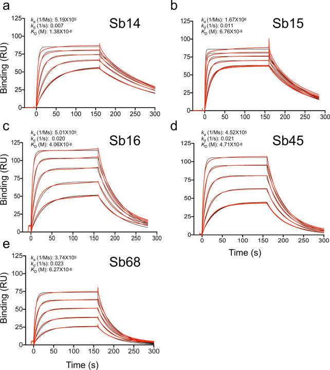
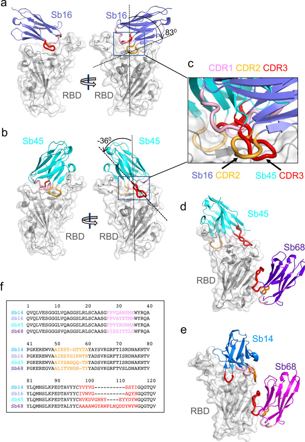
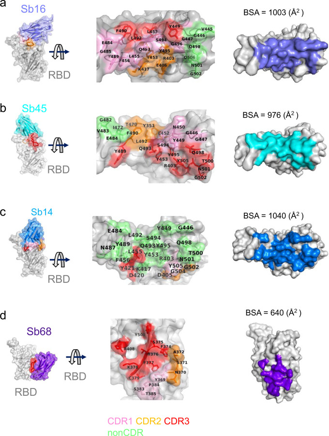
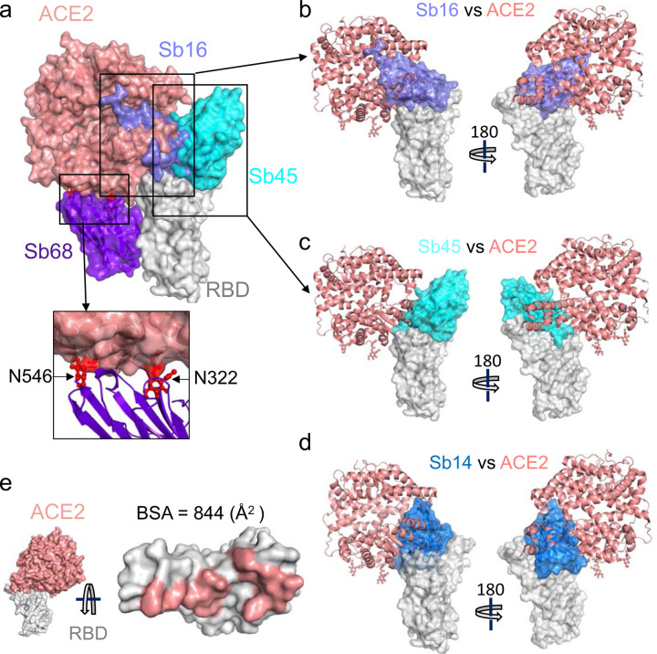
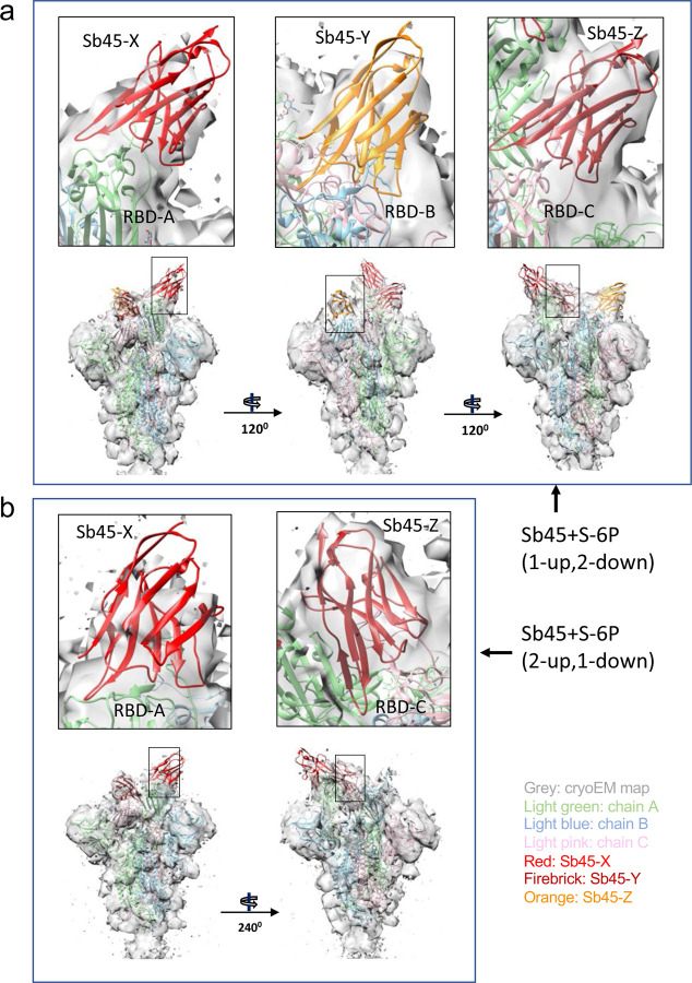
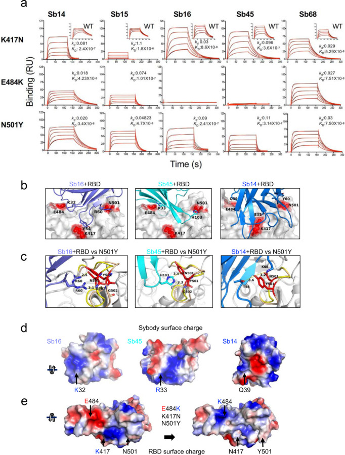
The worldwide spread of severe acute respiratory syndrome coronavirus 2 (SARS-CoV-2) and emergence of new variants demands understanding the structural basis of the interaction of antibodies with the SARS-CoV-2 receptor-binding domain (RBD). Here we report five X-ray crystal structures of sybodies (synthetic nanobodies) including binary and ternary complexes of Sb16-RBD, Sb45-RBD, Sb14-RBD-Sb68, and Sb45-RBD-Sb68; and Sb16 unliganded. These reveal that Sb14, Sb16, and Sb45 bind the RBD at the ACE2 interface and that the Sb16 interaction is accompanied by a large CDR2 shift. In contrast, Sb68 interacts at the periphery of the interface. We also determined cryo-EM structures of Sb45 bound to spike (S). Superposition of the X-ray structures of sybodies onto the trimeric S protein cryo-EM map indicates some may bind both "up" and "down" configurations, but others may not. Sensitivity of sybody binding to several recently identified RBD mutants is consistent with these structures.


Conflict of interest statement

Competing interests. The authors declare no competing interests.

Figures

Fig. 1|
Fig. 1|. Sybodies bind RBD with KD values in the nanomolar range.
RBD was coupled to a biosensor chip as described in Methods. Graded concentrations (31 to 500 nM) of each of the indicated sybodies were offered to the coupled surface (from t=0 to t=160 s), followed by buffer washout, and measurement of net binding (in resonance units, RU). Experimental curves were fit by global analysis using BIAeval 2.0 (Cytiva). Curves shown are representative of at least two determinations.
Fig. 2|
Fig. 2|. Overall structures of Sb14, Sb16, Sb45 and Sb68 complexed with SARS-CoV-2 RBD.
Ribbons (sybodies) and ribbons plus surface (RBD) representations of the complex of (a) Sb16 (slate) with RBD (grey) (7KGK); (b) Sb45 (cyan) with RBD (7KGJ), (d) Sb45 and Sb68 (purple) with RBD (7KLW) and (e) Sb14 (blue) and Sb68 (magenta) with RBD (7MFU). Sb16-RBD and Sb45-RBD superimposed based on the RBD are shown in (c) to highlight CDR loops, which are color coded as CDR1 (pink), CDR2 (orange) and CDR3 (red). The CDR2 of Sb16 and CDR3 of Sb45 interact similarly with the RBD surface. Panel (f) shows a sequence alignment of the four sybodies.
Fig. 3|
Fig. 3|. Interfaces and interactions of sybodies with RBD.
(a) Sb16-RBD, (b) Sb45-RBD, (c) Sb14-RBD, and (d) Sb68-RBD. (Individual contacting residues are listed in Supplementary Table 1). CDR1, CDR2, CDR3 regions are painted pink, orange and red respectively. Additional non-CDR region contacting residues are colored lime. On the RBD surface, the epitopic residues that contact the sybodies are colored according to the sybody CDR.
Fig. 4|
Fig. 4|. Sybodies clash with ACE2 in RBD complex structures.
(a) Sb16 (slate), Sb45 (cyan), Sb14 (blue), and Sb68 (purple) – RBD complexes were superposed on the ACE2–RBD structure (salmon) (6M0J) based on the RBD. Views of Sb16 (b), Sb45 (c), and Sb14 (d) are shown alone as well. Sb14 and Sb16 are buried inside ACE2; Sb45 is partially buried in ACE2; and Sb68 has major clashes with two N-glycan sites (N322 and N546) of ACE2 (inset). (e) Epitopic area (on RBD) captured by ACE2 (salmon) is indicated along with its BSA.
Fig. 5|
Fig. 5|. X-ray model of sybody superposed on cryo-EM Structures of SB45–S-6P.
(a) Model of Sb45+S-6P (1-up, 2-down) is fitted to the map with Sb45-X bound to RBD-A (up), Sb45-Y to RBD-B (down), and Sb45-Z to RBD-C (down), and CC (Sb45-X/Sb45-Y/Sb45-Z) are 0.52/0.49/0.57 respectively; (b) Model of Sb45+S-6P (2-up, 1-down) is fitted to the map with Sb45-X bound to RBD-A (up), and Sb45-Z bound to RBD-C (down), and CC (Sb45-X/Sb45-Z) are 0.47/0.70 respectively.
Fig. 6|
Fig. 6|. RBD mutations affect sybody binding.
(a) SPR binding of each of the indicated sybodies (across top) to each of the individual RBD mutants. Inset shows binding of sybodies to wild type RBD (from Fig. 1). Experimental tracings are shown in red, curve fits in black and kd (s−1) and KD (M) values as determined from global fitting with BIAeval 2.0 are provided in each panel. (b) Location of contacts of Sb16, Sb45, and Sb14 are shown. E484, K417 and N501 of RBD (wild type) interact with K32, Y54 and R60 of Sb16 respectively; E484 and N501 of RBD (wild type) interact with R33 and H103 of Sb45 respectively; and E484, K417 and N501 of RBD (wild type) interact with Q39, E35, and Y60 of Sb14 respectively. (c) Comparison of complex structures with minimized models involving the N501Y mutation. In silico mutagenesis of N501Y was performed using 7KGK (Sb16+RBD), 7KGJ (Sb45+RBD), and 7MFU (Sb14+RBD+Sb68). Following amino acid substitution in Coot, local energy minimization (within 15 to 20 Å of the mutant residue) was performed through three rounds in PHENIX. For the Sb16-RBD complex, when N501 is mutated to Y501, the loop (496–506, from yellow to wheat) extends about 2.4 Å, but R60 (revealing a double conformation) still forms hydrogen bonds with the Y501 loop; for the Sb45-RBD complex, when N501 is mutated to Y501, the loop (496–506, from yellow to wheat) extends about 1.0 Å, but H103 of Sb45 would still interact with Y501; for the Sb14-RBD complex, when N501 is mutated to Y501, the loop (496–506, from yellow to wheat) is extended about 2.0 Å, but T58 and K65 still the hydrogen bonds with Y501; (d) The surface charge of Sb16, K32 forms a hydrogen bond with E484 of RBD with the opposite charge; the surface charge of Sb45, R33 forms a hydrogen bond with E484 of RBD with the opposite charge; the surface charge of Sb14, Q39 (a neutral residue) interacts with E484 of RBD; (e) Surface charge of wild type of RBD and surface charge of RBD with the three mutations (E484, K417N, and N501Y). When E484 is mutated to K484, the surface charge is changed from negative to positive, therefore the hydrogen bonds are broken – pushing Sb16 and Sb45 out of contact, while since Q39 of Sb14 is not a charged residue, it still may interact with K484 of the mutated RBD.

References

    1. Conti P. et al. The British variant of the new coronavirus-19 (Sars-Cov-2) should not create a vaccine problem. J Biol Regul Homeost Agents 35(2021). - PubMed
    1. Kirby T. New variant of SARS-CoV-2 in UK causes surge of COVID-19. Lancet Respir Med (2021). - PMC - PubMed
    1. Tang J.W., Tambyah P.A. & Hui D.S. Emergence of a new SARS-CoV-2 variant in the UK. J Infect (2020). - PMC - PubMed
    1. Wibmer C.K. et al. SARS-CoV-2 501Y.V2 escapes neutralization by South African COVID-19 donor plasma. bioRxiv, 2021.01.18.427166 (2021). - PubMed
    1. Wang Q. et al. Structural and Functional Basis of SARS-CoV-2 Entry by Using Human ACE2. Cell (2020). - PMC - PubMed

Publication types