

COVID-19: Histopathological correlates of imaging patterns on chest computed tomography
- PMID: 34159661
- PMCID: PMC8447040
- DOI: 10.1111/resp.14101
COVID-19: Histopathological correlates of imaging patterns on chest computed tomography
Abstract
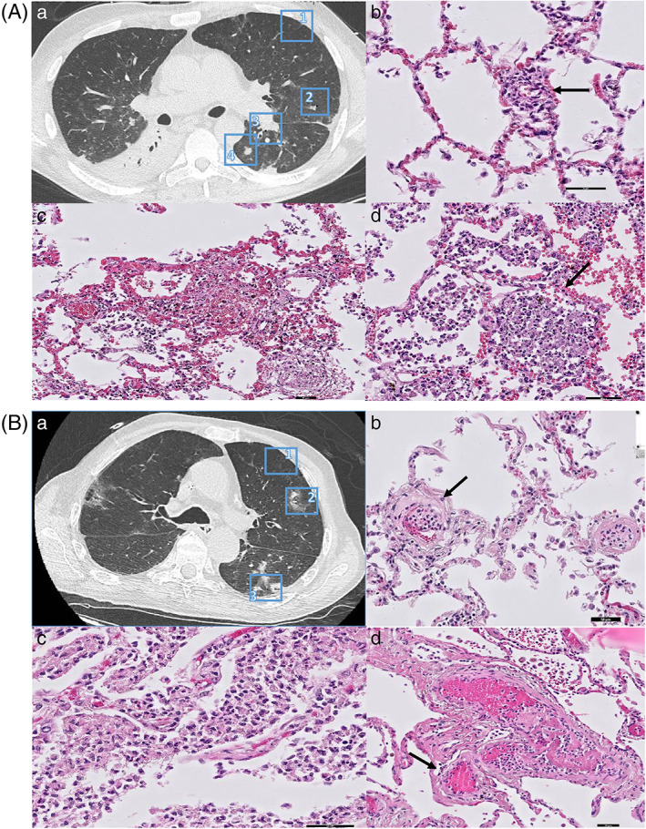
Background and objective: Patients with coronavirus disease 2019 (COVID-19) pneumonia present with typical findings on chest computed tomography (CT), but the underlying histopathological patterns are unknown. Through direct regional correlation of imaging findings to histopathological patterns, this study aimed to explain typical COVID-19 CT patterns at tissue level.
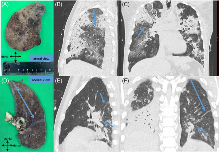
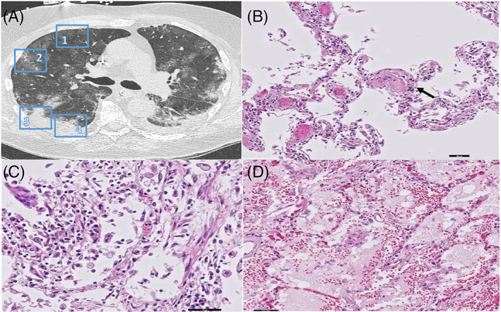
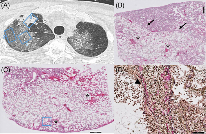
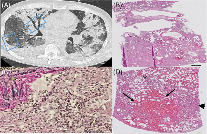
Methods: Eight autopsy cases were prospectively selected of patients with PCR-proven COVID-19 pneumonia with varying clinical manifestations and causes of death. All had been subjected to chest CT imaging 24-72 h prior to death. Twenty-seven lung areas with typical COVID-19 patterns and two radiologically unaffected pulmonary areas were correlated to histopathological findings in the same lung regions.
Results: Two dominant radiological patterns were observed: ground-glass opacity (GGO) (n = 11) and consolidation (n = 16). In seven of 11 sampled areas of GGO, diffuse alveolar damage (DAD) was observed. In four areas of GGO, the histological pattern was vascular damage and thrombosis, with (n = 2) or without DAD (n = 2). DAD was also observed in five of 16 samples derived from areas of radiological consolidation. Seven areas of consolidation were based on a combination of DAD, vascular damage and thrombosis. In four areas of consolidation, bronchopneumonia was found. Unexpectedly, in samples from radiologically unaffected lung parenchyma, evidence was found of vascular damage and thrombosis.
Conclusion: In COVID-19, radiological findings of GGO and consolidation are mostly explained by DAD or a combination of DAD and vascular damage plus thrombosis. However, the different typical CT patterns in COVID-19 are not related to specific histopathological patterns. Microvascular damage and thrombosis are even encountered in the radiologically normal lung.
Keywords: COVID-19; SARS-CoV-2, viral infection; acute respiratory distress syndrome; chest CT; coronavirus disease; histopathological and imaging.
© 2021 The Authors. Respirology published by John Wiley & Sons Australia, Ltd on behalf of Asian Pacific Society of Respirology.
Conflict of interest statement
Prof. Dr Anton Vonk Noordegraaf is supported by the Netherlands CardioVascular Research Initiative (CVON‐2012‐08 PHAEDRA, CVON‐2017‐10 DOLPHIN‐GENESIS) and the Netherlands Organization for Scientific Research (NWO‐VICI: 918.16.610). He also received speaker's money from Johnson & Johnson, and Ferrer in the past 3 years, and served as a member of the scientific advisory board of Acceleron. The other authors report no conflict of interests.
Figures
Comment in
-
Pulmonary vascular involvement in COVID-19 pneumonitis: Is this the first and final insult?Respirology. 2021 Sep;26(9):832-834. doi: 10.1111/resp.14123. Epub 2021 Jul 28. Respirology. 2021. PMID: 34322959 Free PMC article.
Similar articles
-
COVID-19 pneumonia: computer-aided quantification of healthy lung parenchyma, emphysema, ground glass and consolidation on chest computed tomography (CT).Radiol Med. 2021 Apr;126(4):553-560. doi: 10.1007/s11547-020-01305-9. Epub 2020 Nov 18. Radiol Med. 2021. PMID: 33206301 Free PMC article.
-
Comparison of chest radiography and chest CT for evaluation of pediatric COVID-19 pneumonia: Does CT add diagnostic value?Pediatr Pulmonol. 2021 Jun;56(6):1409-1418. doi: 10.1002/ppul.25313. Epub 2021 Feb 25. Pediatr Pulmonol. 2021. PMID: 33631061 Free PMC article.
-
Tracking the time course of pathological patterns of lung injury in severe COVID-19.Respir Res. 2021 Jan 29;22(1):32. doi: 10.1186/s12931-021-01628-9. Respir Res. 2021. PMID: 33514373 Free PMC article.
-
Ground-glass opacity (GGO): a review of the differential diagnosis in the era of COVID-19.Jpn J Radiol. 2021 Aug;39(8):721-732. doi: 10.1007/s11604-021-01120-w. Epub 2021 Apr 26. Jpn J Radiol. 2021. PMID: 33900542 Free PMC article. Review.
-
Postmortem CT pulmonary findings in SARS-CoV-2-positive cases: correlation with lung histopathological findings and autopsy results.Int J Legal Med. 2022 Sep;136(5):1407-1415. doi: 10.1007/s00414-022-02793-2. Epub 2022 Feb 14. Int J Legal Med. 2022. PMID: 35157128 Free PMC article. Review.
Cited by
-
Investigation of the relationship of CO-RADS and CT patterns with laboratory parameters in COVID-19 patients and a new perspective on the total CT scoring system.BMC Med Imaging. 2022 Jul 20;22(1):128. doi: 10.1186/s12880-022-00857-8. BMC Med Imaging. 2022. PMID: 35858851 Free PMC article.
-
Prediction of oxygen supplementation by a deep-learning model integrating clinical parameters and chest CT images in COVID-19.Jpn J Radiol. 2023 Dec;41(12):1359-1372. doi: 10.1007/s11604-023-01466-3. Epub 2023 Jul 13. Jpn J Radiol. 2023. PMID: 37440160 Free PMC article.
-
Respiratory symptoms and radiological findings in post-acute COVID-19 syndrome.ERJ Open Res. 2022 Apr 19;8(2):00479-2021. doi: 10.1183/23120541.00479-2021. eCollection 2022 Apr. ERJ Open Res. 2022. PMID: 35445129 Free PMC article.
-
Characteristic patterns of SARS-CoV-2 on chest CT suggests a hematologic pathway for viral entry into the lung.Clin Imaging. 2022 Sep;89:92-94. doi: 10.1016/j.clinimag.2022.06.011. Epub 2022 Jun 25. Clin Imaging. 2022. PMID: 35772334 Free PMC article.
-
Pulmonary vascular involvement in COVID-19 pneumonitis: Is this the first and final insult?Respirology. 2021 Sep;26(9):832-834. doi: 10.1111/resp.14123. Epub 2021 Jul 28. Respirology. 2021. PMID: 34322959 Free PMC article.
References
-
- Raptis CA, Hammer MM, Short RG, Shah A, Bhalla S, Bierhals AJ, et al. Chest CT and coronavirus disease (COVID‐19): a critical review of the literature to date. AJR Am J Roentgenol. 2020;215:839–42. - PubMed
-
- Goyal N, Chung M, Bernheim A, Keir G, Mei X, Huang M, et al. Computed tomography features of coronavirus disease 2019 (COVID‐19): a review for radiologists. J Thorac Imaging. 2020;35:211–8. - PubMed
Publication types
MeSH terms
Grants and funding
LinkOut - more resources
Full Text Sources
Medical
Miscellaneous
