. 2021 Jun 23;16(6):e0253475.
doi: 10.1371/journal.pone.0253475. eCollection 2021.

Iron overload inhibits BMP/SMAD and IL-6/STAT3 signaling to hepcidin in cultured hepatocytes

Affiliations

Iron overload inhibits BMP/SMAD and IL-6/STAT3 signaling to hepcidin in cultured hepatocytes

Edouard Charlebois et al. PLoS One. .

Abstract

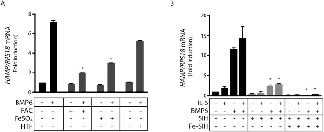
Hepcidin is a peptide hormone that targets the iron exporter ferroportin, thereby limiting iron entry into the bloodstream. It is generated in hepatocytes mainly in response to increased body iron stores or inflammatory cues. Iron stimulates expression of bone morphogenetic protein 6 (BMP6) from liver sinusoidal endothelial cells, which in turn binds to BMP receptors on hepatocytes and induces the SMAD signaling cascade for transcriptional activation of the hepcidin-encoding HAMP mRNA. SMAD signaling is also essential for inflammatory HAMP mRNA induction by the IL-6/STAT3 pathway. Herein, we utilized human Huh7 hepatoma cells and primary murine hepatocytes to assess the effects of iron perturbations on signaling to hepcidin. Iron chelation appeared to slightly impair signaling to hepcidin. Subsequent iron supplementation not only failed to reverse these effects, but drastically reduced basal HAMP mRNA and inhibited HAMP mRNA induction by BMP6 and/or IL-6. Thus, treatment of cells with excess iron inhibited basal and BMP6-mediated SMAD5 phosphorylation and induction of HAMP, ID1 and SMAD7 mRNAs in a dose-dependent manner. Iron also inhibited IL-6-mediated STAT3 phosphorylation and induction of HAMP and SOCS3 mRNAs. These responses were accompanied by induction of GCLC and HMOX1 mRNAs, known markers of oxidative stress. We conclude that hepatocellular iron overload suppresses hepcidin by inhibiting the SMAD and STAT3 signaling pathways downstream of their respective ligands.


Conflict of interest statement

The authors have declared that no competing interests exist.

Figures

Fig 1
Fig 1. Iron overload inhibits SMAD and STAT signaling downstream of BMP6 and IL-6 in Huh7 hepatoma cells.
Huh 7 cells were treated with 100 μM DFO for 18 hours before washing and supplementation with 50 μM FAC. After 2 hours, cells were treated with 20 ng/ml human IL-6, 5 ng/ml BMP6, or both over 4 hours. (A) qPCR analysis of HAMP mRNA. (B) Western blot analysis of pSTAT3, STAT3, pSMAD5, SMAD5, ferritin and β-actin. (C-F) qPCR analysis of ID1, TFRC, GCLC and HMOX1 mRNAs. All data in graphs are presented as the mean ± SEM from three independent experiments for (A) and two independent experiments for (B-F). Statistical analysis was performed by one-way ANOVA. Statistically significant differences (p<0.05) across iron treatments (compared to respective values from iron-unperturbed cells shown in black bars) are indicated by *.
Fig 2
Fig 2. Iron salts but not transferrin-bound iron inhibit hepcidin induction by BMP6 and/or IL-6 in Huh7 hepatoma cells.
(A) Huh 7 cells were pretreated with either 50 μM FAC, 50 μM FeSO4, or 30 μM holo-transferrin (HTF) for 2 hours and then treated with BMP6 for 4 hours; HAMP mRNA was measured by qPCR. (B) Huh 7 cells were treated with 100 μM SIH for 18 hours before washing and supplementation with 50 Fe-SIH. After 2 hours, cells were treated with 20 ng/ml IL-6, 5ng/ml BMP6, or both over 4 hours. HAMP mRNA was measured by qPCR. All data in graphs are presented as the mean ± SEM from three independent experiments. Statistical analysis was performed by one-way ANOVA. Statistically significant differences (p<0.05) across iron treatments (compared to respective values from iron-unperturbed cells shown in black bars) are indicated by *.
Fig 3
Fig 3. Dose-dependent inhibitory effects of iron on basal and BMP6-mediated SMAD signaling and hepcidin expression in Huh7 hepatoma cells.
Huh7 cells were treated with increasing doses of FAC over 2 hours. When indicated, the experiment was either terminated or the cells were washed and further incubated with 25 ng/ml BMP6 (A and C) or 5 ng/ml BMP6 (B, D-F) for 4 hours. (A and B) qPCR analysis of HAMP and ID1 mRNAs. (C) Western blot analysis of pSMAD5, SMAD5 and β-actin. (D-F) qPCR analysis of HAMP, ID1 and GCLC mRNAs. All data in graphs are presented as the mean ± SEM from three independent experiments. Statistical analysis was performed by one-way ANOVA (A, B, D and E) two-way ANOVA (F). Statistically significant differences (p<0.05) across iron treatments (compared to respective values from iron-unperturbed cells shown in black bars) are directly shown or indicated by *.
Fig 4
Fig 4. Iron overload inhibits Smad and Stat signaling downstream of BMP6 and IL-6 in primary murine hepatocytes.
Primary murine hepatocytes were maintained in serum-free William’s Medium E over the course of experiments. The cells were pretreated with 100 μM DFO for 18 hours and then washed and supplemented with 50 μM FAC over 2 hours. The cells were then treated with 20 ng/ml murine IL-6, 25 ng/ml BMP6, or both over 4 hours. (A) qPCR analysis of Hamp mRNA. (B) Western blotting of pSTAT3, STAT3, pSMAD5, SMAD1, ferritin, and β-actin. (C-F) qPCR analysis of Id1, Smad7, Socs3 and Tfrc mRNAs. All data in graphs are presented as the mean ± SEM from two independent experiments. Statistical analysis was performed by one-way ANOVA. Statistically significant differences (p<0.05) across iron treatments (compared to respective values from iron-unperturbed cells shown in black bars) are indicated by *.

References

    1. Ganz T. Systemic iron homeostasis. Physiol Rev. 2013;93(4):1721–41. Epub 2013/10/19. doi: 10.1152/physrev.00008.2013 . - DOI - PubMed
    1. Wang CY, Babitt JL. Liver iron sensing and body iron homeostasis. Blood. 2019;133(1):18–29. Epub 2018/11/08. doi: 10.1182/blood-2018-06-815894 . - DOI - PMC - PubMed
    1. Coffey R, Ganz T. Iron homeostasis: An anthropocentric perspective. J Biol Chem. 2017;292(31):12727–34. Epub 2017/06/16. doi: 10.1074/jbc.R117.781823 . - DOI - PMC - PubMed
    1. Ganz T, Nemeth E. Iron homeostasis in host defence and inflammation. Nat Rev Immunol. 2015;15(8):500–10. doi: 10.1038/nri3863 . - DOI - PMC - PubMed
    1. Katsarou A, Pantopoulos K. Basics and principles of cellular and systemic iron homeostasis. Mol Aspects Med. 2020;75:100866. Epub 2020/06/23. doi: 10.1016/j.mam.2020.100866 . - DOI - PubMed

Publication types

Grants and funding