. 2021 Jun 22;35(12):109205.
doi: 10.1016/j.celrep.2021.109205.

DHX15 is required to control RNA virus-induced intestinal inflammation

Affiliations

DHX15 is required to control RNA virus-induced intestinal inflammation

Junji Xing et al. Cell Rep. .

Abstract

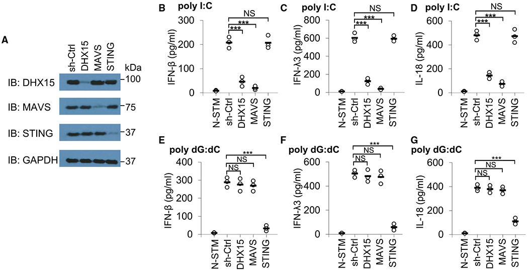
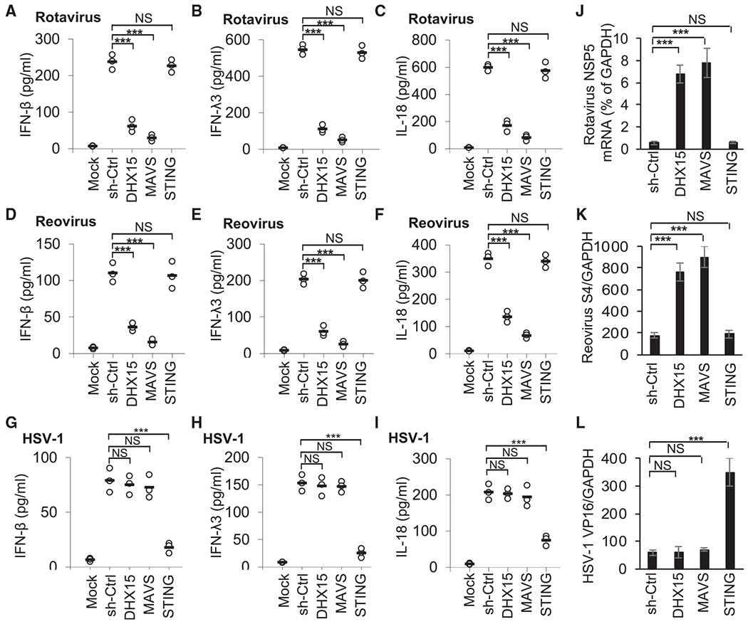
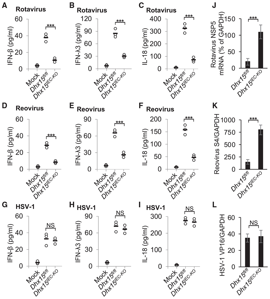
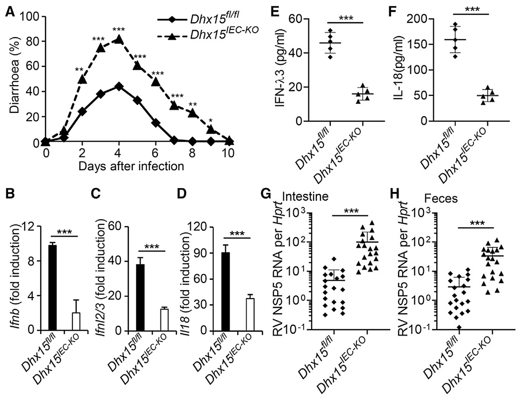
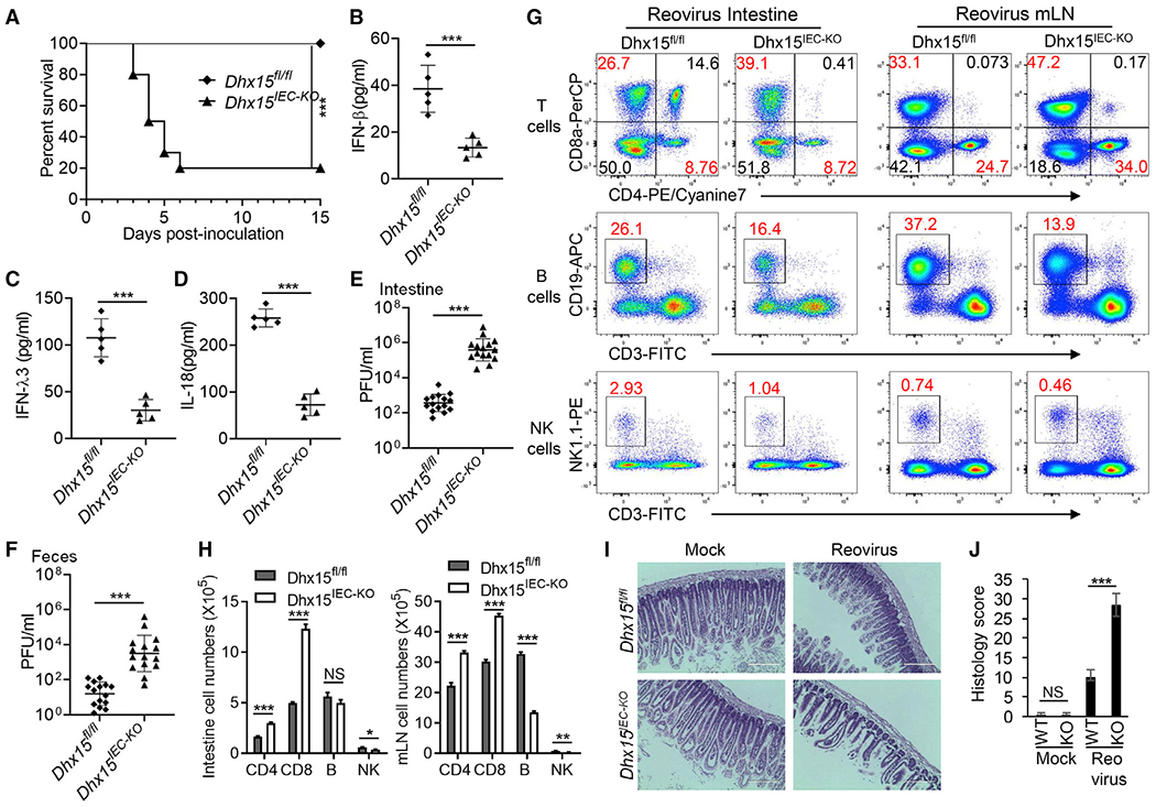
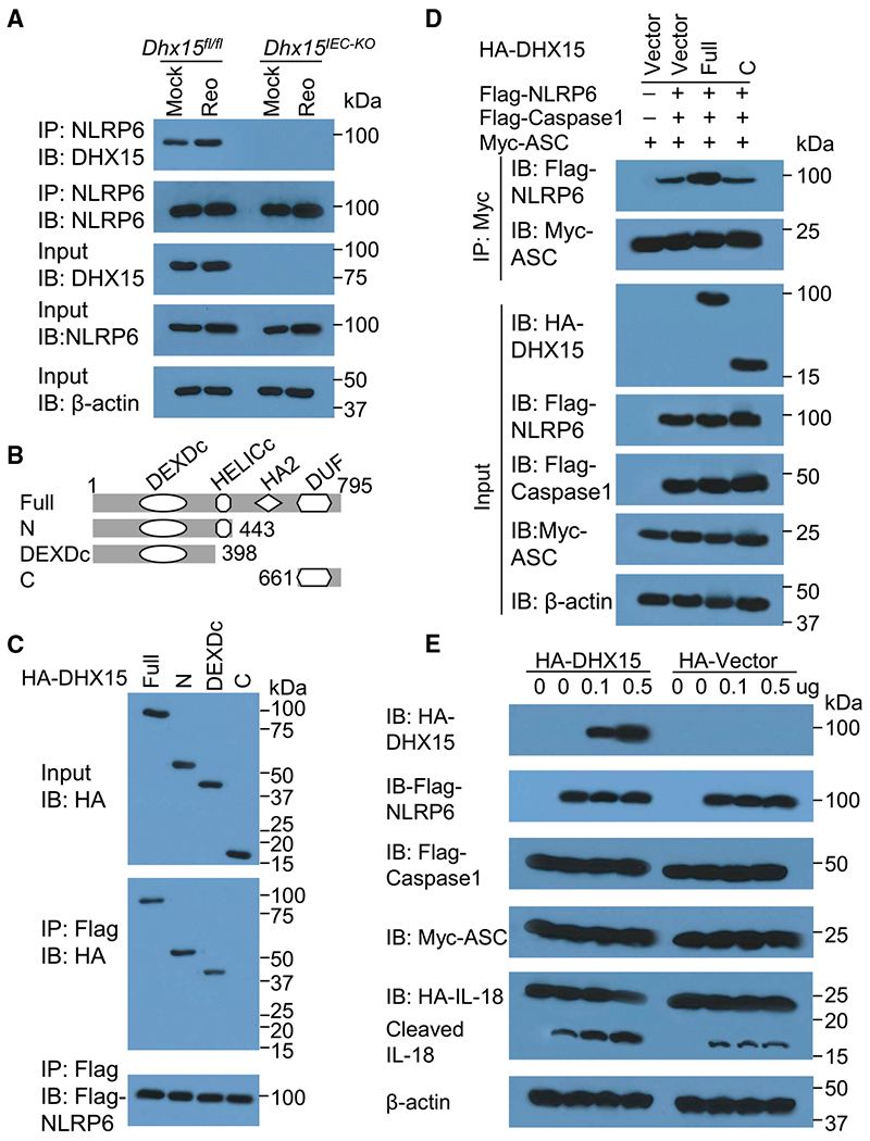
RNA helicases play critical roles in various biological processes, including serving as viral RNA sensors in innate immunity. Here, we find that RNA helicase DEAH-box helicase 15 (DHX15) is essential for type I interferon (IFN-I, IFN-β), type III IFN (IFN-λ3), and inflammasome-derived cytokine IL-18 production by intestinal epithelial cells (IECs) in response to poly I:C and RNA viruses with preference of enteric RNA viruses, but not DNA virus. Importantly, we generate IEC-specific Dhx15-knockout mice and demonstrate that DHX15 is required for controlling intestinal inflammation induced by enteric RNA virus rotavirus in suckling mice and reovirus in adult mice in vivo, which owes to impaired IFN-β, IFN-λ3, and IL-18 production in IECs from Dhx15-deficient mice. Mechanistically, DHX15 interacts with NLRP6 to trigger NLRP6 inflammasome assembly and activation for inducing IL-18 secretion in IECs. Collectively, our report reveals critical roles for DHX15 in sensing enteric RNA viruses in IECs and controlling intestinal inflammation.


Conflict of interest statement

Declaration of interests The authors declare no competing interests.

Figures

Figure 1.
Figure 1.. DHX15 is essential for producing IFN-β, IFN-λ3, and IL-18 by human HT-29 IECs in response to poly I:C
(A) Immunoblot (IB) showing the knockdown efficiency of shRNAs targeting the indicated genes in HT-29 IECs. Nontargeting shRNA served as a control (sh-Ctrl). Glyceraldehyde 3-phosphate dehydrogenase (GAPDH) blots are shown as loading controls. The position of protein markers (shown in kDa) is indicated at right. (B–G) ELISA of IFN-β (B and E), IFN-λ3 (C and F), and IL-18 (D and G) production from human HT-29 IECs with the indicated shRNA after a 20-h stimulation with 5 μg/mL poly I:C (B–D) or 2.5 μg/mL poly dG:dC (E–G) delivered by Lipofectamine 3000. N-STM, scrambled shRNA-treated HT-29 IECs without stimulation. Each circle represents an individual independent experiment, and small solid black lines indicate the average of triplicates. NS, not significant; p > 0.05, ***p < 0.001 (unpaired t test).
Figure 2.
Figure 2.. DHX15 is required for IFN-β, IFN-λ3, and IL-18 production in human HT-29 IECs upon enteric RNA virus infection
(A–I) ELISA of IFN-β (A, D, and G), IFN-λ3 (B, E, and H), and IL-18 (C, F, and I) production from human HT-29 IECs with the indicated shRNA after a 20-h infection with enteric RNA viruses, including simian rotavirus SA-11 strain (A–C) and reovirus T3D strain (D–F), or DNA virus HSV-1 KOS strain (G–I) at a multiplicity of infection (MOI) of 10. Mock, scrambled shRNA (sh-Ctrl)-treated human HT-29 IECs without virus infection. Each circle represents an individual independent experiment, and small solid black lines indicate the average of triplicates. (J–L) Quantification of expression of rotavirus NSP5 gene (J), reovirus S4 gene (K), and HSV-1 VP16 gene (L) relative to GAPDH in human HT-29 IECs infected by rotavirus (J), reovirus (K), or HSV-1(L) as in (A)–(I). Data are represented as means ± SEMs. NS, p > 0.05, ***p < 0.001 (unpaired t test).
Figure 3.
Figure 3.. DHX15 positively regulates production of IFN-β, IFN-λ3, and IL-18 in mouse primary IECs upon enteric RNA virus infection
(A–I) ELISA of IFN-β (A, D, and G), IFN-λ3 (B, E, and H) and IL-18 (C, F, and I) production in mouse primary IECs from wild-type Dhx15fl/fl and Dhx15IEC-KO mice after a 20-h infection with enteric RNA viruses, including rotavirus EW strain (A–C) and reovirus T3D strain (D–F), or DNA virus HSV-1 KOS strain (G–I) at a MOI of 10. Mock, cells without virus infection. Each circle represents an individual independent experiment, and small solid black lines indicate the average of triplicates. (J–L) Quantification of expression of rotavirus NSP5 gene (J), reovirus S4 gene (K), and HSV-1 VP16 gene (L) relative to GAPDH in mouse primary IECs from wild-type Dhx15fl/fl and Dhx15IEC-KO mice infected by rotavirus (J), reovirus (K), or HSV-1(L) as in (A)–(I). Data are represented as means ± SEMs. NS, p > 0.05, ***p < 0.001 (unpaired t test).
Figure 4.
Figure 4.. DHX15 is essential to control intestinal inflammation induced by enteric rotavirus infection in suckling mice in vivo
(A) Diarrhea duration and percentage of mice with diarrhea (score ≥ 2) from 8-day-old wild-type Dhx15fl/fl and Dhx15IEC-KO suckling mice (n = 20 per strain) orally inoculated by gavage with 1 DD50 rotavirus EW strain. (B–F) The wild-type Dhx15fl/fl and Dhx15IEC-KO suckling mice (n = 5 per strain) were orally inoculated by gavage with 1 DD50 rotavirus EW strain. At day 1 post-inoculation, mice were euthanized, and intestine tissues were excised for qRT-PCR detection of Ifnb (B), Ifnl2/3 (C), and Il18 (D) expression. In addition, the excised intestine was homogenized in PBS for the detection of IFN-λ3 (E) and IL-18 (F) in intestine homogenates by ELISA. Data are represented as means ± SEMs. (G and H) The wild-type Dhx15fl/fl and Dhx15IEC-KO suckling mice (n = 20 per strain) were orally inoculated by gavage with 1 DD50 rotavirus EW strain. At day 5 post-inoculation, mice were euthanized, and intestine tissues (G) and feces (H) were collected for qRT-PCR detection of rotavirus levels. Mock, mouse without rotavirus infection. *p < 0.05, **p < 0.01, and ***p < 0.001 (unpaired t test).
Figure 5.
Figure 5.. DHX15 is required for control of intestinal inflammation induced by enteric reovirus infection in adult mice in vivo
(A) Survival of 5-week-old wild-type Dhx15fl/fl and Dhx15IEC-KO adult mice (n = 10 per strain) after intragastric injection of reovirus T3D strain (1 × 10⁸ plaque-forming units [PFUs] per mouse). (B–D) The wild-type Dhx15fl/fl and Dhx15IEC-KO mice (n = 5 per strain) were inoculated intragastrically with 1 × 10⁸ PFUs reovirus T3D strain. At day 1 post-inoculation, mice were euthanized, and intestine tissues were excised and homogenized in PBS. Levels of IFN-β (B), IFN-λ3 (C), and IL-18 (D) in intestine homogenates were quantified by ELISA. (E and F) The wild-type Dhx15fl/fl and Dhx15IEC-KO mice (n = 15 per strain) were inoculated intragastrically with 1 × 10⁸ PFU of reovirus T3D strain. At day 4 post-inoculation, mice were euthanized, feces were collected, and intestinal tissues were excised. The viral titers in intestine homogenates (E) and shedding in feces (F) were determined by plaque assay. Results are expressed as mean viral titers for 15 animals for each time point. Error bars indicate SEMs. (G) Flow cytometry analysis of CD4+ T cells, CD8+ T cells, B cells, and NK cells of intestine lamina propria lymphocytes (left panel) and mesenteric lymph nodes (mLN) (right panel) from wild-type Dhx15fl/fl and Dhx15IEC-KO mice infected with reovirus for 2 days using CD3-FITC, CD4-PE/cyanine7, CD8a-PerCP/cyanine5.5, CD19-APC, and NK1.1-PE antibodies. (H) The absolute cell numbers in intestine (left) and mLN (right) from wild-type Dhx15fl/fl and Dhx15IEC-KO mice (n = 3 mice) for representative flow cytometry data in (G). (I) Hematoxylin and eosin (H&E) staining of intestine sections from wild-type Dhx15fl/fl and Dhx15IEC-KO mice as in (E). Scale bars represent 200 μm. (J) Graph depicting histology scores for inflammation and tissue damage of intestine sections in (I). Data are represented as means ± SEMs. NS, p > 0.05, *p < 0.05, **p < 0.01, and ***p < 0.001 (unpaired t test).
Figure 6.
Figure 6.. DHX15 recruits NLRP6 to promote the inflammasome assembly and activation
(A) IB analysis of endogenous proteins of DHX15 and NLRP6 precipitated with anti-NLRP6 from whole-cell lysates of IECs from wild-type Dhx15fl/fl and Dhx15IEC-KO mice infected without (Mock) or with reovirus (Reo) T3D strain at MOI of 5. (B) Schematic diagram showing full-length (Full) DHX15 and its truncations with deletion of various domains (left margin); numbers at ends indicate amino acid positions (top). DEXDc, DEAD-like helicases superfamily domain; HELICc, helicase superfamily c-terminal domain; HA2, helicase-associated domain 2; DUF, domain of unknown function. (C) IB analysis of purified FLAG-tagged NLRP6 with anti-FLAG (bottom blot), and IB analysis (with anti-HA) of purified HA-tagged full-length DHX15 (Full) and DHX15 truncation mutants alone (top blot) including N-terminal domain (N), DEAD-like helicases superfamily domain (DEXDc), and C-terminal domain (C), or after incubation with FLAG-tagged NLRP6 and immunoprecipitation with anti-FLAG (center blot). (D) IB analysis of DHX15, NLRP6, caspase-1, and ASC in HEK293T cells transfected with Myc-ASC and co-transfected with or without FLAG-caspase-1, FLAG-NLRP6, HA-DHX15 (Full for full-length, or C for its truncate containing C-terminal domain), or HA-vector control followed by immunoprecipitation with anti-Myc antibody. (E) IB analysis of DHX15, NLRP6, caspase-1, ASC, full-length IL-18, and its cleaved IL-18 in HEK293T cells transfected with HA-IL-18, Myc-ASC, and FLAG-caspase-1, and co-transfected with or without FLAG-NLRP6, HA-DHX15, or HA-vector control with different doses as indicated. The position of protein markers (shown in kDa) is indicated at right.

References

    1. Ablasser A, and Hur S (2020). Regulation of cGAS- and RLR-mediated immunity to nucleic acids. Nat. Immunol 21, 17–29. - PubMed
    1. Ablasser A, Goldeck M, Cavlar T, Deimling T, Witte G, Röhl I, Hopfner KP, Ludwig J, and Hornung V (2013). cGAS produces a 2′-5′-linked cyclic dinucleotide second messenger that activates STING. Nature 498, 380–384. - PMC - PubMed
    1. Angel J, Franco MA, Greenberg HB, and Bass D (1999). Lack of a role for type I and type II interferons in the resolution of rotavirus-induced diarrhea and infection in mice. J. Interferon Cytokine Res 19, 655–659. - PubMed
    1. Atarashi K, Tanoue T, Shima T, Imaoka A, Kuwahara T, Momose Y, Cheng G, Yamasaki S, Saito T, Ohba Y, et al. (2011). Induction of colonic regulatory T cells by indigenous Clostridium species. Science 331, 337–341. - PMC - PubMed
    1. Ball JM, Tian P, Zeng CQ, Morris AP, and Estes MK (1996). Age-dependent diarrhea induced by a rotaviral nonstructural glycoprotein. Science 272, 101–104. - PubMed

Publication types