Clinical Trial
. 2021 Jul 22;184(15):3949-3961.e11.
doi: 10.1016/j.cell.2021.06.002. Epub 2021 Jun 5.

The monoclonal antibody combination REGEN-COV protects against SARS-CoV-2 mutational escape in preclinical and human studies

Affiliations
Clinical Trial

The monoclonal antibody combination REGEN-COV protects against SARS-CoV-2 mutational escape in preclinical and human studies

Richard Copin et al. Cell. .

Abstract

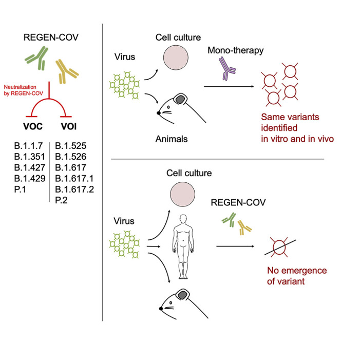
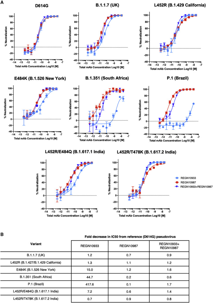
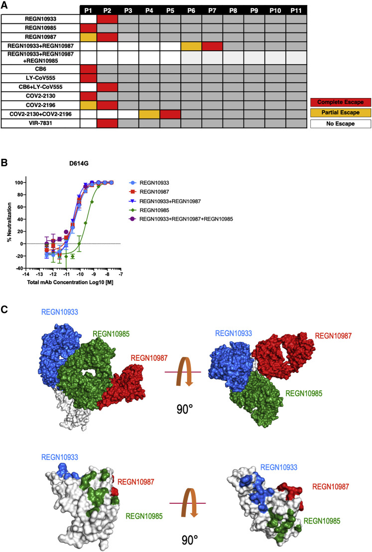
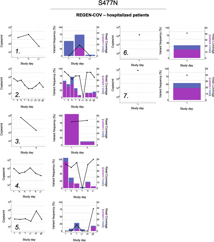
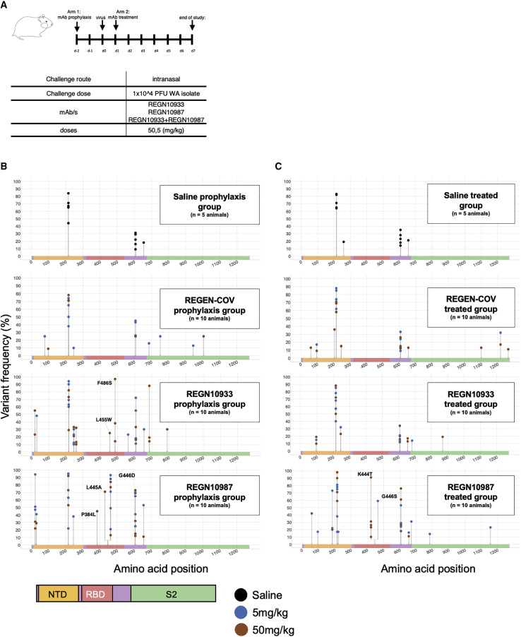
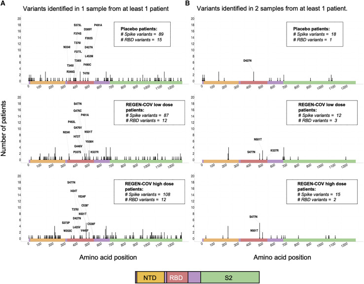
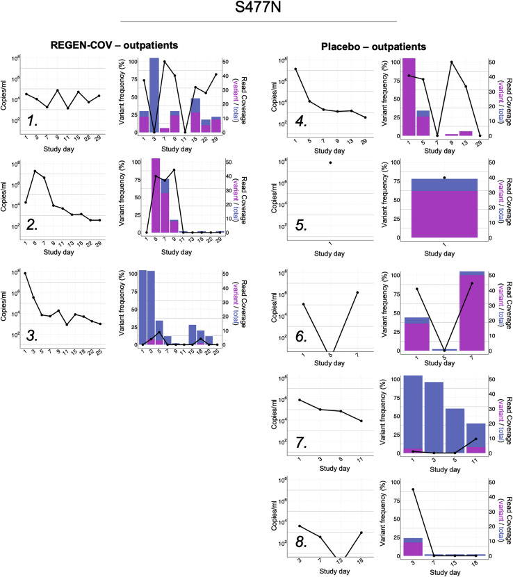
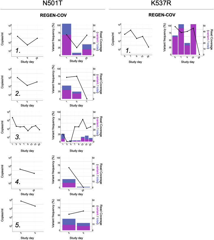
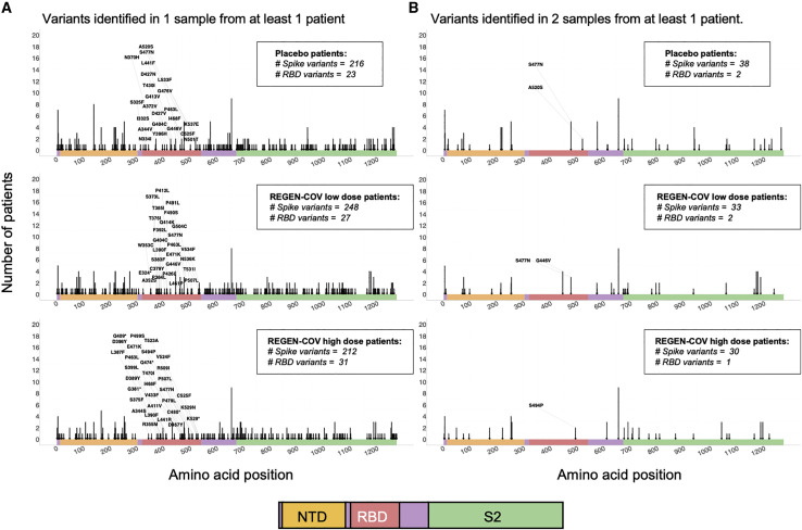
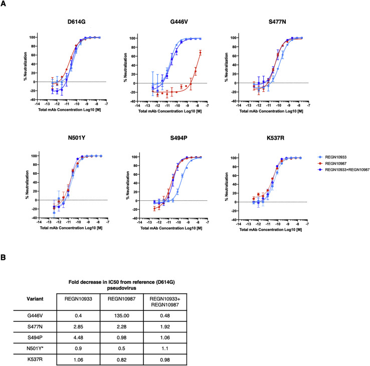
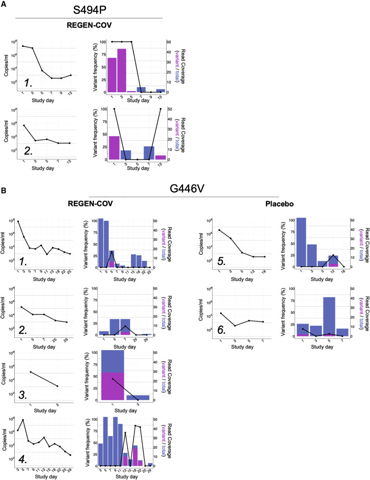
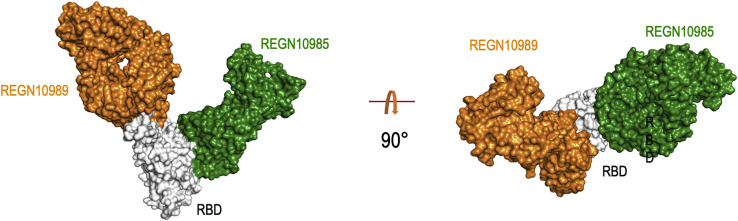
Monoclonal antibodies against SARS-CoV-2 are a clinically validated therapeutic option against COVID-19. Because rapidly emerging virus mutants are becoming the next major concern in the fight against the global pandemic, it is imperative that these therapeutic treatments provide coverage against circulating variants and do not contribute to development of treatment-induced emergent resistance. To this end, we investigated the sequence diversity of the spike protein and monitored emergence of virus variants in SARS-COV-2 isolates found in COVID-19 patients treated with the two-antibody combination REGEN-COV, as well as in preclinical in vitro studies using single, dual, or triple antibody combinations, and in hamster in vivo studies using REGEN-COV or single monoclonal antibody treatments. Our study demonstrates that the combination of non-competing antibodies in REGEN-COV provides protection against all current SARS-CoV-2 variants of concern/interest and also protects against emergence of new variants and their potential seeding into the population in a clinical setting.

Keywords: COVID-19; REGEN-COV; SARS-CoV-2; escape; monoclonal antibodies.


Conflict of interest statement

Declaration of interests Regeneron authors own options and/or stock of the company. This work has been described in one or more pending provisional patent applications. G.S.A., G.H., D.M.W., L.L., N.S., A.J.M., G.D.Y., and C.A.K. are officers of Regeneron.

Figures

None
Graphical abstract
Figure 1
Figure 1
Neutralization of variants of concern and variants of interest by the REGEN-COV combination and individual antibodies Neutralization curves and (A) Neutralization fold-decrease (B) relative to parental D614G variant. Full UK B.1.1.7 (H69del, V70del, Y145del, N501Y, A570D, D614G, P681H, T716I, S982A, D1118H), South African B.1.351 (D80Y, D215Y, L241del, L242del, A243del, L242del, K417N, E484K, N501Y, D614G, A701V) and P.1 (L18F, T20N, P26S, D138Y, R190S, K417T, E484K, N501Y, D614G, H655Y, T1027I, V1176F) variants were assessed. Key RBD residues from B.1.429 California (L452R) and B.1.617.1 India (L452R/E484Q) and B.1.617.2 India (L452R/T478K) variants were assessed. The E484K variant has been identified in many global lineages, including the B.1.526 New York, P.2 Brazil and B.1.618 India lineages. Data represent three technical replicates.
Figure 2
Figure 2
In vitro escape with individual antibodies and antibody combinations VSV-spike recombinant virus was passaged under pressure of individual antibodies or antibody combinations. (A) Passage number during which the virus completely escaped neutralizing activity of the antibodies as evidenced by the cytopathic effect (CPE) is shown as “complete escape” for wells where antibody concentration was 10 μg/mL or above or “partial escape” for wells where antibody concentration was 1–10 μg/mL. (B) Neutralization potency of the two mAb REGN10987+REGN10933 combination and the three mAb REGN10987+REGN10933+REGN10987+REGN10985 combination along with individual mAbs. Data represent three technical replicates. (C) Two views of the RBD-REGN10933-REGN10987-REGN10985 model. RBD is colored white; REGN10933 is shown blue, REGN10987 is shown in red and REGN10985 is shown in green. Surface representation of the RBD in white with contact patches for each antibody, colors as in (A). See also Figure S4 and Table S1.
Figure S1
Figure S1
Cryo-EM structure of RBD-REGN10989-REGN10985 in surface representation, related to Figure 2 RBD is in white, REGN10985 in green and REGN19089 in orange.
Figure 3
Figure 3
Frequency of SARS-CoV-2 spike variants identified in viruses from lungs of hamsters treated with either REGN10987 or REGN10933 as monotherapy or with the REGEN-COV combination in both prophylactic and treatment settings (A) Study design (n = 5 for placebo group, n = 10 for treatment groups). (B and C) Graphs show the distribution and frequencies of amino acid variants across the spike protein sequence identified in samples from the (B) prophylactic or (C) treated groups. Mutated sites are indicated with gray lines and dots show the frequency of variants in each animal. The color of each dot indicated the dose used. Spike protein domain is color coded as indicated, and all amino acid changes in RBD are labeled. Study represents one biological replicate. See also Table S3.
Figure 4
Figure 4
Variant analysis of virus sequences in SARS-CoV-2-positive samples from hospitalized patients Graphs show the distribution of amino acid variants across the spike protein sequence identified in samples from the placebo, REGEN-COV low dose-, and REGEN-COV high dose-treated patients. Mutated sites are indicated with arrows, and arrow length designates the number of patients with 1 or more variant-containing samples. Spike protein domains are color coded as indicated and all amino acid changes in RBD are labeled. (A) Arrows highlight variants identified in 1 or more samples from at least one patient. (B) Arrows highlight variants identified in 2 or more samples from at least one patient. All samples were characterized by the defining glycine substitution at position 614.
Figure S2
Figure S2
Viral load and longitudinal frequencies of S477N in SARS-CoV-2-positive samples from hospitalized patients, related to Figure 5 Viral load was determined by quantitative RT-PCR. Variant frequencies are indicated by the black line as percent of reads with nSNPs relative to total number of covering reads. Histograms show total (blue) and mutated (magenta) read coverage at variant position.
Figure 5
Figure 5
Viral load and longitudinal frequencies of variants in REGN-COV2-treated, SARS-CoV-2-positive samples from hospitalized patients Viral load was determined by quantitative RT-PCR. N501T (A) and K537R (B) variant frequencies are indicated by the black line as percent of reads with nSNPs relative to total number of covering reads. Histograms show total (blue) and mutated (magenta) read coverage at variant position. See also Figures S1 and S3 and Tables S2 and S4.
Figure 6
Figure 6
Variant analysis of virus sequences in SARS-CoV-2-positive samples from non-hospitalized patients Graphs show the distribution of amino acid variants across the spike protein sequence identified in samples from the placebo, REGEN-COV low dose-, and REGEN-COV high dose-treated patients. Mutated sites are indicated with arrows, and arrow length designates the number of patients with 1 or more variant-containing samples. Spike protein domains are color coded as indicated and all amino acid changes in RBD are labeled. (A) Arrows highlight variants identified in 1 or more samples from at least one patient. (B) Arrows highlight variants identified in 2 or more samples from at least one patient. All samples were characterized by the defining glycine substitution at position 614. See also Figures S2 and S3 and Tables S2 and S4.
Figure S3
Figure S3
Viral load and longitudinal frequencies of S477N in SARS-CoV-2-positive samples from non-hospitalized patients, related to Figure 6 Viral load was determined by quantitative RT-PCR. Variant frequencies are indicated by the black line as percent of reads with nSNPs relative to total number of covering reads. Histograms show total (blue) and mutated (magenta) read coverage at variant position.
Figure 7
Figure 7
Viral load and longitudinal frequencies of variants in SARS-CoV-2-positive samples from non-hospitalized patients Viral load was determined by quantitative RT-PCR. S494P (A) and G446V (B) variant frequencies are indicated by the black line as percent of reads with nSNPs relative to total number of covering reads. Histograms show total (blue) and mutated (magenta) read coverage at variant position. See also Tables S2 and S4.
Figure S4
Figure S4
Clinical variant neutralization by the REGEN-COV combination and individual antibodies, related to Figures 5 and 6 Variants identified in two or more samples from at least one patient in either the hospitalized or outpatient trial were assessed for impact on neutralization potency. A) Neutralization curves and B) Fold-change decrease versus parental D614G virus. The N501Y variant was used to substitute for N501T.

References

    1. Baum A., Fulton B.O., Wloga E., Copin R., Pascal K.E., Russo V., Giordano S., Lanza K., Negron N., Ni M., et al. Antibody cocktail to SARS-CoV-2 spike protein prevents rapid mutational escape seen with individual antibodies. Science. 2020;369:1014–1018. - PMC - PubMed
    1. Baum A., Ajithdoss D., Copin R., Zhou A., Lanza K., Negron N., Ni M., Wei Y., Mohammadi K., Musser B., et al. REGN-COV2 antibodies prevent and treat SARS-CoV-2 infection in rhesus macaques and hamsters. Science. 2020;370:1110–1115. - PMC - PubMed
    1. Centers for Disease Control and Prevention . 2021. SARS-CoV-2 Variant Classifications and Definitions.https://www.cdc.gov/coronavirus/2019-ncov/variants/variant-info.html
    1. Davies N.G., Jarvis C.I., Group C.C., -19 W., Edmunds W.J., Jewell N.P., Diaz-Ordaz K., Keogh R.H. Increased mortality in community-tested cases of SARS-CoV-2 lineage B.1.1.7. medRxiv. 2021 doi: 10.1101/2021.02.01.21250959. - DOI - PMC - PubMed
    1. Dong J., Zost S.J., Greaney A.J., Starr T.N., Dingens A.S., Chen E.C., Chen R.E., Case J.B., Sutton R.E., Gilchuk P., et al. Genetic and structural basis for recognition of SARS-CoV-2 spike protein by a two-antibody cocktail. bioRxiv. 2021 doi: 10.1101/2021.01.27.428529. - DOI - PMC - PubMed

Publication types

Substances