

Oral wound healing models and emerging regenerative therapies
- PMID: 34161876
- PMCID: PMC8380729
- DOI: 10.1016/j.trsl.2021.06.003
Oral wound healing models and emerging regenerative therapies
Abstract
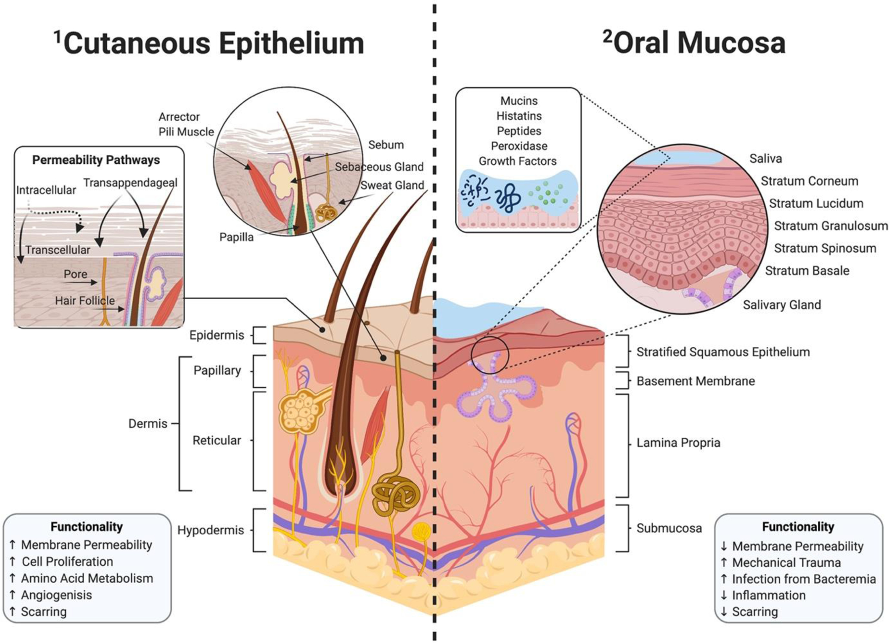
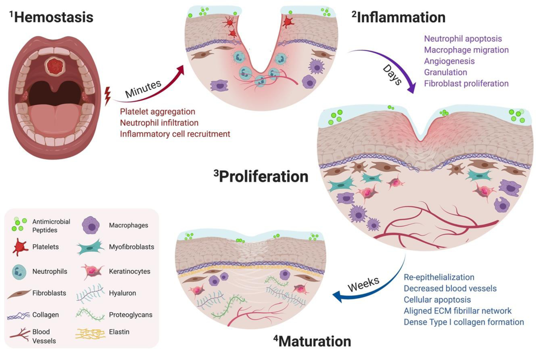
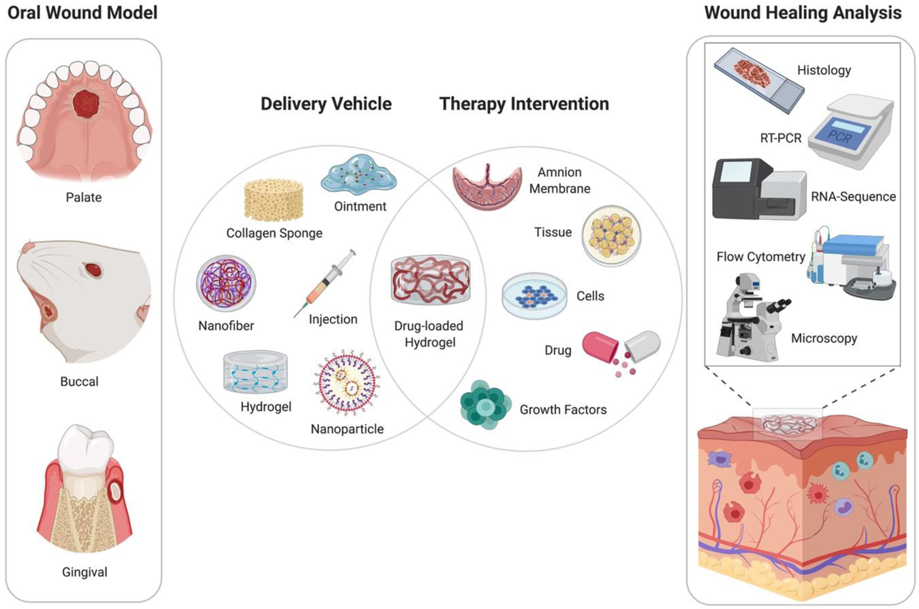
Following injury, the oral mucosa undergoes complex sequences of biological healing processes to restore homeostasis. While general similarities exist, there are marked differences in the genomics and kinetics of wound healing between the oral cavity and cutaneous epithelium. The lack of successful therapy for oral mucosal wounds has influenced clinicians to explore alternative treatments and potential autotherapies to enhance intraoral healing. The present in-depth review discusses current gold standards for oral mucosal wound healing and compares endogenous factors that dictate the quality of tissue remodeling. We conducted a review of the literature on in vivo oral wound healing models and emerging regenerative therapies published during the past twenty years. Studies were evaluated by injury models, therapy interventions, and outcome measures. The success of therapeutic approaches was assessed, and research outcomes were compared based on current hallmarks of oral wound healing. By leveraging therapeutic advancements, particularly within in cell-based biomaterials and immunoregulation, there is great potential for translational therapy in oral tissue regeneration.
Copyright © 2021 The Authors. Published by Elsevier Inc. All rights reserved.
Figures
Similar articles
-
Development and preclinical evaluation of acellular collagen scaffolding and autologous artificial connective tissue in the regeneration of oral mucosa wounds.Tissue Eng Part A. 2010 May;16(5):1667-79. doi: 10.1089/ten.TEA.2008.0571. Tissue Eng Part A. 2010. PMID: 20001832
-
Biomimetic hydrogel for rapid and scar-free healing of skin wounds inspired by the healing process of oral mucosa.Acta Biomater. 2019 Dec;100:255-269. doi: 10.1016/j.actbio.2019.10.011. Epub 2019 Oct 10. Acta Biomater. 2019. PMID: 31606531
-
An overview of the therapeutic potential of regenerative medicine in cutaneous wound healing.Int Wound J. 2017 Jun;14(3):450-459. doi: 10.1111/iwj.12735. Epub 2017 Mar 6. Int Wound J. 2017. PMID: 28261962 Free PMC article. Review.
-
Improving hard palate wound healing using immune modulatory autotherapies.Acta Biomater. 2019 Jun;91:209-219. doi: 10.1016/j.actbio.2019.04.052. Epub 2019 Apr 25. Acta Biomater. 2019. PMID: 31029828 Free PMC article.
-
Wound healing and regenerative strategies.Oral Dis. 2011 Sep;17(6):541-9. doi: 10.1111/j.1601-0825.2011.01787.x. Epub 2011 Feb 18. Oral Dis. 2011. PMID: 21332599 Review.
Cited by
-
The Oral Wound Healing Potential of Thai Propolis Based on Its Antioxidant Activity and Stimulation of Oral Fibroblast Migration and Proliferation.Evid Based Complement Alternat Med. 2022 May 26;2022:3503164. doi: 10.1155/2022/3503164. eCollection 2022. Evid Based Complement Alternat Med. 2022. PMID: 35664934 Free PMC article.
-
Attached Oral Mucosal Wound Closure using Blue Glue - A Prospective Clinical Study.Ann Maxillofac Surg. 2023 Jan-Jun;13(1):31-36. doi: 10.4103/ams.ams_2_23. Epub 2023 Jul 28. Ann Maxillofac Surg. 2023. PMID: 37711540 Free PMC article.
-
Immediate but Temporal Response: The Role of Distal Epithelial Cells in Wound Healing.Stem Cell Rev Rep. 2024 Aug;20(6):1587-1598. doi: 10.1007/s12015-024-10734-2. Epub 2024 May 17. Stem Cell Rev Rep. 2024. PMID: 38760627 Free PMC article.
-
Magnesium metal-organic framework microneedles loaded with curcumin for accelerating oral ulcer healing.J Nanobiotechnology. 2024 Sep 30;22(1):594. doi: 10.1186/s12951-024-02873-y. J Nanobiotechnology. 2024. PMID: 39350179 Free PMC article.
-
Oral Wound Healing Potential of Polygoni Cuspidati Rhizoma et Radix Decoction-In Vitro Study.Pharmaceuticals (Basel). 2023 Feb 10;16(2):267. doi: 10.3390/ph16020267. Pharmaceuticals (Basel). 2023. PMID: 37259412 Free PMC article.
References
-
- Liu J., et al., Skin and oral mucosa equivalents: construction and performance. Orthod Craniofac Res, 2010. 13(1): p. 11–20. - PubMed
-
- Squier CA and Kremer MJ, Biology of oral mucosa and esophagus. J Natl Cancer Inst Monogr, 2001(29): p. 7–15. - PubMed
-
- Arda O, Goksugur N, and Tuzun Y, Basic histological structure and functions of facial skin. Clin Dermatol, 2014. 32(1): p. 3–13. - PubMed
-
- Losquadro WD, Anatomy of the Skin and the Pathogenesis of Nonmelanoma Skin Cancer. Facial Plast Surg Clin North Am, 2017. 25(3): p. 283–289. - PubMed
Publication types
MeSH terms
Grants and funding
LinkOut - more resources
Full Text Sources
Miscellaneous
