. 2021 Jun 24;18(1):142.
doi: 10.1186/s12974-021-02178-z.

Divanillyl sulfone suppresses NLRP3 inflammasome activation via inducing mitophagy to ameliorate chronic neuropathic pain in mice

Affiliations

Divanillyl sulfone suppresses NLRP3 inflammasome activation via inducing mitophagy to ameliorate chronic neuropathic pain in mice

Shuai Shao et al. J Neuroinflammation. .

Abstract

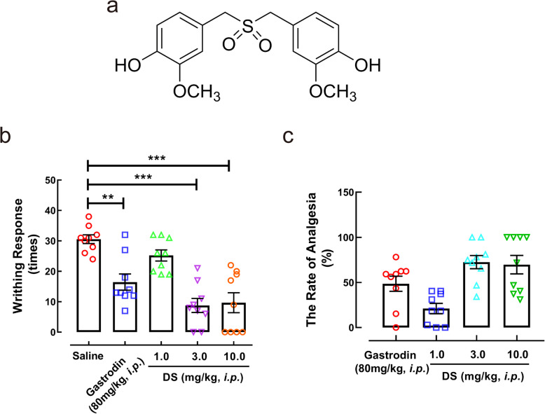
Background: Chronic neuropathic pain is a frequent sequel to peripheral nerve injury and maladaptive nervous system function. Divanillyl sulfone (DS), a novel structural derivative of 4,4'-dihydroxydibenzyl sulfoxide from a traditional Chinese medicine Gastrodia elata with anti-nociceptive effects, significantly alleviated neuropathic pain following intrathecal injection. Here, we aimed to investigate the underlying mechanisms of DS against neuropathic pain.

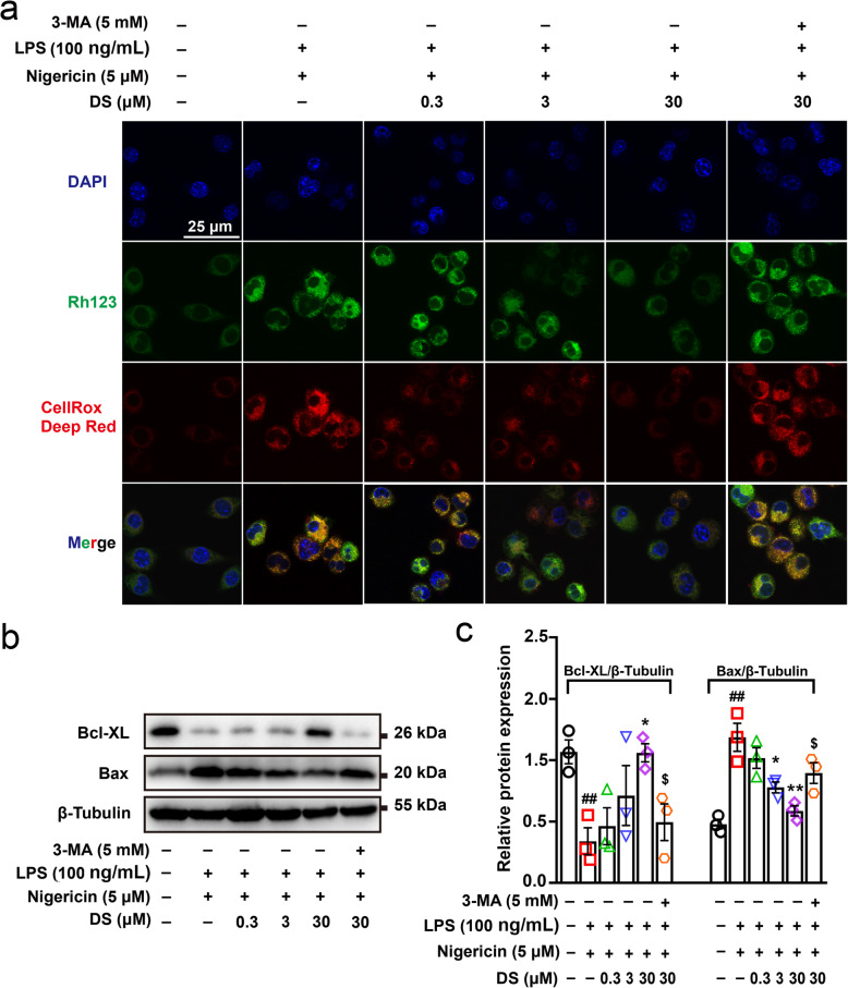
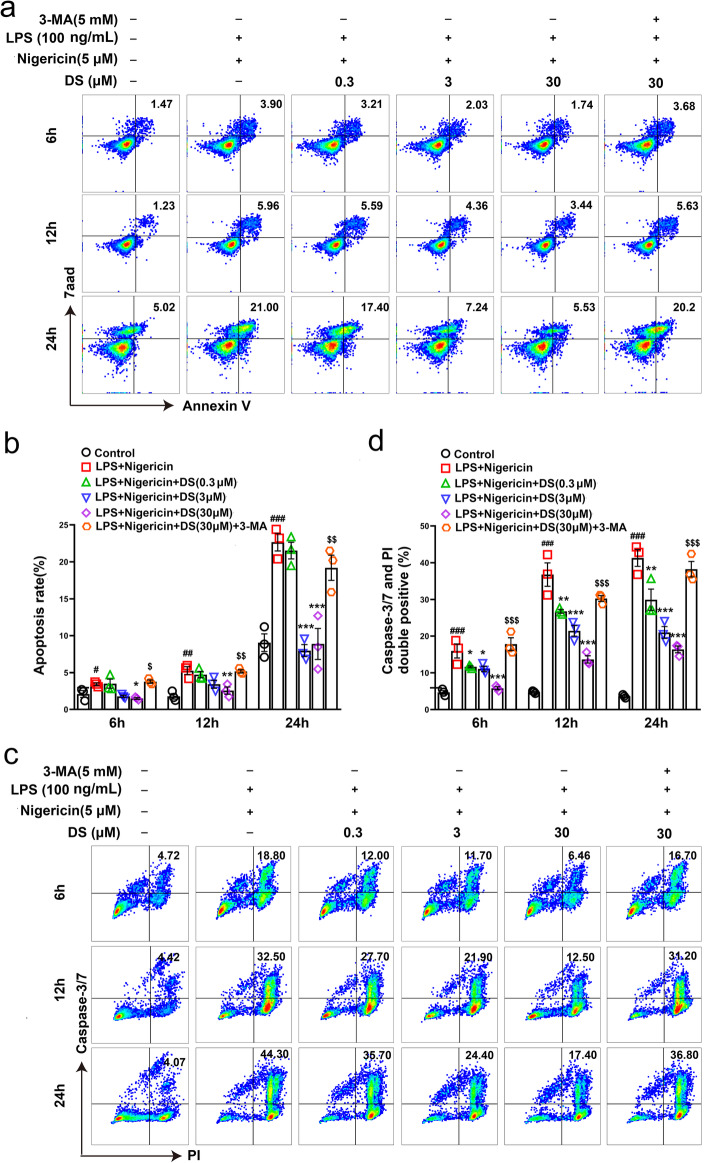
Methods: A chronic constrictive injury (CCI) mouse model of neuropathic pain induced by sciatic nerve ligation was performed to evaluate the effect of DS by measuring the limb withdrawal using Von Frey filament test. Immunofluorescence staining was used to assess the cell localizations and expressions of Iba-1, ASC, NLRP3, and ROS, the formation of autolysosome. The levels of NLRP3-related proteins (caspase-1, NLRP3, and IL-1β), mitophagy-related proteins (LC3, Beclin-1, and p62), and apoptosis-related proteins (Bcl-XL and Bax) were detected by Western blotting. The apoptosis of BV-2 cell and caspase activity were evaluated by flow cytometry.

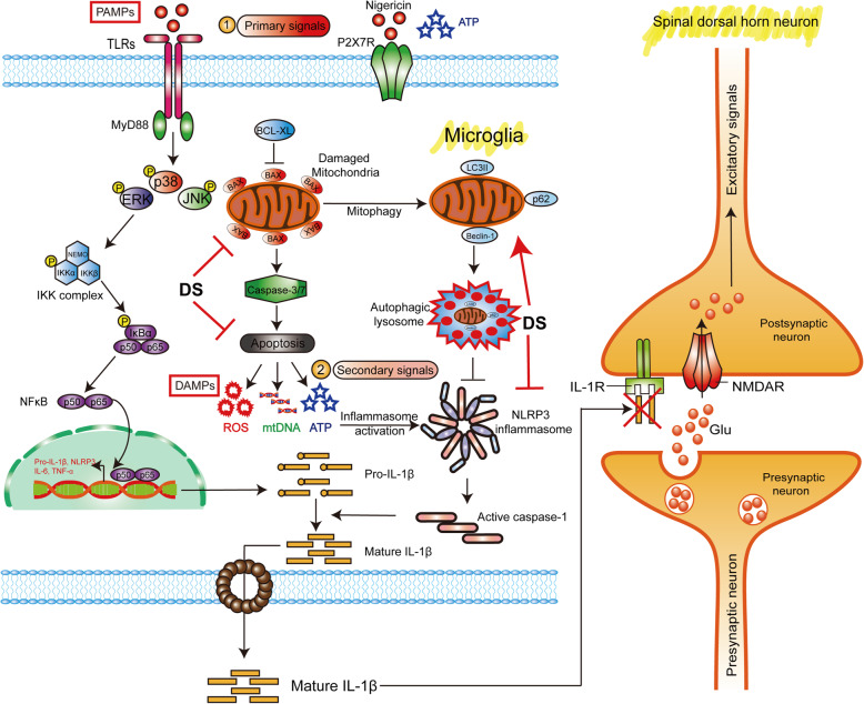
Results: DS significantly alleviated the neuropathic pain by increasing the mechanical withdrawal threshold and inhibiting the activation of NLRP3 in CCI-induced model mice. Our findings indicated that DS promoted the mitophagy by increasing the LC3II and Beclin 1 and decreasing the levels of p62 protein in BV-2 cell. This is accompanied by the inhibition of NLRP3 activation, which was shown as inhibited the expression of NLRP3 in lysates as well as the secretion of mature caspase-1 p10 and IL-1β p17 in supernatants in cultured BV-2 microglia. In addition, DS could promote mitophagy-induced improvement of dysfunctional mitochondria by clearing intracellular ROS and restoring mitochondrial membrane potential.

Conclusion: Together, our findings demonstrated that DS ameliorate chronic neuropathic pain in mice by suppressing NLRP3 inflammasome activation induced by mitophagy in microglia. DS may be a promising therapeutic agent for chronic neuropathic pain.

Keywords: Chronic neuropathic pain; Divanillyl sulfone; Microglia; Mitophagy; NLRP3 inflammasome.


Conflict of interest statement

The authors declare that they have no competing interests.

Figures

Fig. 1
Fig. 1
DS attenuated pain hypersensitivity of somatic pain in vivo. a The chemical structure of DS. DS significantly decreased the writhing times (b) and increased the rate of analgesia (c) in acetic acid-induced mice. Data are expressed as mean ± SEM (n = 9 mice in each group). Statistical significance was determined by one-way ANOVA followed by Tukey’s post hoc analysis where **P < 0.01, ***P < 0.001 vs. saline group
Fig. 2
Fig. 2
DS treatment improved sciatic nerve ligation-induced chronic neuropathic pain. Neuropathic pain mice were induced by ligation of sciatic nerve, the mechanical withdrawal threshold within 14 days after surgery (a) and intrathecal injection of vehicle (0.9% normal saline) or DS (1, 3, 10 mg/kg) 9 days after surgery (b) were measured by Von Frey Monofilament in both contralateral and ipsilateral paws. c The analgesic effects of both drugs over four consecutive days post-injection. d Dose-response analysis of DS on paw withdrawal threshold 0.5 h after administration, and ED50 was calculated with GraphPad Prism 8 with normalized to maximal possible effect. Data are expressed as means ± SEM (n = 8 mice in each group). Statistical significance was determined by two-way ANOVA followed by Tukey’s post hoc analysis where *P < 0.05, **P < 0.01, ***P < 0.001 vs. CCI + Saline group, ***P < 0.001 vs. Sham-ipsilateral paw group
Fig. 3
Fig. 3
DS showed analgesic effect by inhibiting NLRP3 inflammasome activation of spinal microglia in vivo. a The immunofluorescence of the spinal lumbar enlargement of CCI pain mice showed the co-localization of IBA-1 staining (red) and NLRP3 staining (green) in the ipsilateral glial area, and the contralateral side was not affected. b, c Western blotting analysis of microglia activation marker IBA-1 and NLRP3 inflammasome components in spinal lumbar enlargement tissue showed that DS inhibited the expression of IBA-1, NLRP3 protein, activated caspase-1 p10, and mature IL-1β p17 in a dose-dependent manner. Data are expressed as means ± SEM (n = 3 in each group). Statistical significance was determined by one-way ANOVA followed by Tukey’s post hoc analysis where ##P < 0.01, ###P < 0.011 vs. Sham + Saline group; *P < 0.05, **P < 0.01, ***P < 0.001 vs. CCI + Saline group. NLRP3 inflammasome inhibitor MCC950 (d) and IL-1R antagonist (e) were injected intrathecally into CCI pain mice and the paw withdrawal threshold of their ipsilateral and contralateral paws were measured at 1h post intrathecal injection. Inhibiting the assembly and activation of NLRP3 inflammasome or block the binding of IL-1β to IL-1R can effectively increase the paw withdrawal threshold of CCI mice. Data are expressed as means ± SEM (n = 8 in each group). Statistical significance was determined by two-way ANOVA followed by Tukey’s post hoc analysis where **P < 0.01, ***P < 0.001 vs. CCI + Saline group
Fig. 4
Fig. 4
DS inhibited NLRP3 inflammasome activation and promoted mitophagy in microglia in vitro. BV-2 microglia were first pre-stimulated by LPS (100 ng/ml) for 3.5 h, then treated for 1h with indicated concentrations of DS followed by stimulation with nigericin (5 μM) for 30 min. a, b Immunoblot analyses of caspase-1 p10, IL-1β p17 in culture supernatants (SN) and NLRP3, pro-caspase-1, pro-IL-1β in lysates (LYS) were shown. c The level of IL-1β in supernatants was analyzed by ELISA method. After pre-stimulated with LPS, adding autophagy inhibitor 3-MA (5 mM) before DS and incubated for 0.5 h, and finally stimulated with nigericin for 12 h. d, e Immunoblot analyses of NLRP3, LC3II/I, Beclin 1 and p62 in LYS and c ELISA analysis of IL-1β in SN. Data are expressed as means ± SEM (n = 3 in each group). Statistical significance was determined by one-way ANOVA followed by Tukey’s post hoc analysis where ##P < 0.01, ###P < 0.001 vs. control; *P < 0.05, **P < 0.01, ***P < 0.001 vs. LPS + Nigericin group; $P < 0.05, $$P < 0.01, vs. LPS + Nigericin + 30 μM DS group. e Mitochondria and lysosome were respectively labeled with the mito-tracker (green) and lyso-tracker (red), images were captured using Leica TCS SP8 confocal microscope
Fig. 5
Fig. 5
DS negatively regulated the formation of NLRP3 inflammasome complex through mitophagy in BV-2 microglia. BV-2 microglia were first pre-stimulated by LPS (100 ng/ml) for 3.5 h, then treated for 1h with indicated concentrations of DS and 3-MA (5 mM) for 30 min or not followed by stimulation with nigericin (5 μM) for 6 h, 12 h, and 24 h, which was labeled with a triangle at nigericin (5 μM) treatment. a Immunoblot analyses of caspase-1 p10, IL-1β p17 in culture supernatants (SN) and NLRP3, pro-caspase-1, pro-IL-1β in lysates (LYS) were shown. b Immunofluorescence staining show the expression of NLRP3 (green) and ASC (Red) in BV-2 microglia after stimulation with nigericin for 12 h. c, d Caspase-1 activity was detected by flow cytometry in BV-2 microglia treated with DS for 12 h using caspase-1/PI double-labeled staining method. Data are expressed as means ± SEM (n = 3 in each group). Statistical significance was determined by one-way ANOVA followed by Tukey’s post hoc analysis where ###P < 0.001 vs. control group; ***P < 0.001 vs. LPS + Nigericin group; $$$P < 0.001 vs. LPS + Nigericin + 30 μM DS group
Fig. 6
Fig. 6
DS promoted autophagic clearance of mitochondrial ROS and protected MMP through mitophagy in microglia. BV-2 microglia were first pre-stimulated by LPS (100 ng/ml) for 3.5 h, then treated for 1 h with indicated concentrations of DS and 3-MA (5 mM) for 30 min or not followed by stimulation with nigericin (5 μM) for 12 h. a Immunofluorescence staining Rh123 (green) and CellROX Deep Red (Red) indicate mitochondrial membrane depolarization and ROS production in BV-2 microglia. b, c Immunoblot analyses the expression of apoptosis associated proteins of Bcl-XL and Bax in lysates. Data are expressed as means ± SEM (n = 3 in each group). Statistical significance was determined by one-way ANOVA followed by Tukey’s post hoc analysis where ##P < 0.01 vs. control group; *P < 0.05, **P < 0.01 vs. LPS + Nigericin group; $P < 0.05 vs. LPS + Nigericin + 30 μM DS group
Fig. 7
Fig. 7
DS inhibited apoptosis and caspase-3/7 activity through mitophagy in BV-2 microglia. BV-2 microglia were first pre-stimulated by LPS (100 ng/ml) for 3.5 h, then treated for 1 h with indicated concentrations of DS and 3-MA (5 mM) for 30 min or not followed by stimulation with nigericin (5 μM) for 12 h. Apoptosis was detected by flow cytometry in BV-2 microglia treated with indicated concentrations of DS for 6, 12, and 24 h using Annexin V-FITC/7aad (a, b) and caspase-3/7/PI (c, d) double-labeled staining method. Data are expressed as means ± SEM (n = 3 in each group). Statistical significance was determined by one-way ANOVA followed by Tukey’s post hoc analysis where #P < 0.05, ##P < 0.01, ###P < 0.001 vs. control group; *P < 0.05, **P < 0.01, ***P < 0.001 vs. LPS + Nigericin group; $P < 0.05, $$P < 0.01, $$$P < 0.001 vs. LPS + Nigericin + 30 μM DS group
Fig. 8
Fig. 8
DS ameliorated neuropathic pain through mitophagy-mediated NLRP3 inflammasome inhibition. a Paw withdrawal threshold was measured by Von Frey test in both contralateral and ipsilateral paws, after administrating DS (10 mg/kg), DS (10 mg/kg) + 3-MA (2.5 mg/kg), and vehicle (0.9% normal saline) 9 days in CCI-induced model mice, respectively. b The analgesic effects of both DS (10 mg/kg) and DS (10 mg/kg) + 3-MA (2.5 mg/kg) treatment over four consecutive days post-injection. Data are expressed as means ± SEM (n = 7 mice in each group). Statistical significance was determined by two-way ANOVA followed by Tukey’s post hoc analysis where **P < 0.01 vs. CCI + Saline group, #P < 0.05, ##P < 0.01, ###P < 0.001 vs. CCI + 10 mg/kg DS group. c, d Western blotting analysis of NLRP3 inflammasome components in spinal lumbar enlargement tissue. Data are expressed as means ± SEM (n = 3 in each group). Statistical significance was determined by one-way ANOVA followed by Tukey’s post hoc analysis where #P < 0.05, ##P < 0.01 vs. Sham + Saline group; *P < 0.05 vs. CCI + Saline group; $P < 0.05 vs. CCI + 10 mg/kg DS group
Fig. 9
Fig. 9
Proposed analgesic mechanism of DS for mitophagy-induced NLRP3 inflammasome inhibition in spinal microglia

References

    1. Zeilhofer HU, Benke D, Yevenes GE. Chronic pain states: pharmacological strategies to restore diminished inhibitory spinal pain control. Annu Rev Pharmacol Toxicol. 2012;52(1):111–133. doi: 10.1146/annurev-pharmtox-010611-134636. - DOI - PubMed
    1. Bahari Z, Meftahi GH. Spinal α -adrenoceptors and neuropathic pain modulation; therapeutic target. Br J Pharmacol. 2019;176(14):2366–2381. doi: 10.1111/bph.14580. - DOI - PMC - PubMed
    1. Peirs C, Seal RP. Neural circuits for pain: recent advances and current views. Science. 2016;354(6312):578–584. doi: 10.1126/science.aaf8933. - DOI - PMC - PubMed
    1. Gao YJ, Ji RR. Chemokines, neuronal-glial interactions, and central processing of neuropathic pain. Pharmacol Ther. 2010;126(1):56–68. doi: 10.1016/j.pharmthera.2010.01.002. - DOI - PMC - PubMed
    1. Tanga FY, Nutile-McMenemy N, DeLeo JA. The CNS role of toll-like receptor 4 in innate neuroimmunity and painful neuropathy. Proc Natl Acad Sci U S A. 2005;102(16):5856–5861. doi: 10.1073/pnas.0501634102. - DOI - PMC - PubMed

MeSH terms