. 2021 Jun 29;118(26):e2100474118.
doi: 10.1073/pnas.2100474118.

Lateral gate dynamics of the bacterial translocon during cotranslational membrane protein insertion

Affiliations

Lateral gate dynamics of the bacterial translocon during cotranslational membrane protein insertion

Evan Mercier et al. Proc Natl Acad Sci U S A. .

Abstract

During synthesis of membrane proteins, transmembrane segments (TMs) of nascent proteins emerging from the ribosome are inserted into the central pore of the translocon (SecYEG in bacteria) and access the phospholipid bilayer through the open lateral gate formed of two helices of SecY. Here we use single-molecule fluorescence resonance energy transfer to monitor lateral-gate fluctuations in SecYEG embedded in nanodiscs containing native membrane phospholipids. We find the lateral gate to be highly dynamic, sampling the whole range of conformations between open and closed even in the absence of ligands, and we suggest a statistical model-free approach to evaluate the ensemble dynamics. Lateral gate fluctuations take place on both short (submillisecond) and long (subsecond) timescales. Ribosome binding and TM insertion do not halt fluctuations but tend to increase sampling of the open state. When YidC, a constituent of the holotranslocon, is bound to SecYEG, TM insertion facilitates substantial opening of the gate, which may aid in the folding of YidC-dependent polytopic membrane proteins. Mutations in lateral gate residues showing in vivo phenotypes change the range of favored states, underscoring the biological significance of lateral gate fluctuations. The results suggest how rapid fluctuations of the lateral gate contribute to the biogenesis of inner-membrane proteins.

Keywords: YidC; membrane proteins; ribosome; single-molecule biophysics; translocon SecYEG.


Conflict of interest statement

The authors declare no competing interest.

Figures

Fig. 1.
Fig. 1.
Single-molecule FRET labels to monitor lateral gate dynamics by TIRF. (A) Models of SecYEG in the closed (3J45.pdb) and open (3J00.pdb) conformations were used for coarse-grained simulations of donor (Cy3, green) and acceptor (Atto647N, red) fluorophores attached to cysteine side chains at positions 298 and 148 of SecY. The modeled accessible volumes predict an average distance between donor (D) and acceptor (A) of 31 Å (closed) or 54 Å (open). (B, Upper) Representative fluorescence trace from a single particle upon donor excitation. Donor fluorescence is plotted in light green and acceptor fluorescence in light red. The dark lines are idealized fits of the donor and acceptor traces. a.u., arbitrary units. (B, Lower) FRET trace computed from the donor and acceptor fluorescence in Upper. The red line represents the idealized FRET trace obtained from HMM.
Fig. 2.
Fig. 2.
smFRET histograms of SecYEG fitted with four-state models. (A) FRET histogram obtained from immobilized nanodisc-reconstituted SecYEG complexes measured in TIRF. The blue curve indicates the cumulative distribution comprised of four Gaussian functions (red) obtained from fitting with σ = 0.1. (Inset) Mean FRET values and populations of FRET states along with SDs. (B) Same distribution as A fitted with a four-state HMM (purple). (C) Two-dimensional histogram of FRET efficiency vs. stoichiometry. Stoichiometry (S) is defined as S = nD/(nD + nA) for each single molecule, where nD is the number of donor fluorophores and nA is the number of acceptor fluorophores. (D) FRET histogram obtained from freely diffusing nanodisc-reconstituted SecYEG complexes measured by PIE-FRET. The blue curve indicates the cumulative distribution composed of four Gaussian functions (red) obtained from fitting. (Inset) Mean FRET values and populations of FRET states. (E) Relative populations of the four FRET states calculated from the histograms in A, B, and D. FRET states from low to high FRET are labeled open (green), partially open (blue), partially closed (orange), and closed (red).
Fig. 3.
Fig. 3.
Kinetic analysis of lateral gate opening by HMM. (A) Transition density plot indicating the average FRET before and after each transition identified by four-state HMM analysis. (B) Associated kinetic mechanism.
Fig. 4.
Fig. 4.
PIE-FRET analysis of freely diffusing nanodisc-reconstituted SecYEG. (A) FRET histograms were computed after dividing each burst into time windows of fixed length (colored lines). The raw FRET histogram is plotted for comparison (gray). (B) BVA of the PIE-FRET data were performed by computing the SD of FRET, σ(FRET), for each burst (contour) and averaged over specific FRET intervals (blue symbols). The gray shaded area represents the 99.9% confidence interval computed assuming static FRET states.
Fig. 5.
Fig. 5.
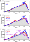
Effect of ligand binding on smFRET of SecYEG. (A) Overlay of FRET histograms obtained for SecYEG alone (gray area), with 70S ribosomes (blue), or with SRP receptor, FtsY (red). The peak range for each dataset, calculated by Monte Carlo simulation, is indicated by a solid bar above the histogram. Significant differences compared to SecYEG alone are indicated (**P < 0.001). (B) Same as A but with LepB75-RNC (red), LepB94-RNC (blue), or AqpZ75-RNC (purple). (C) Same as A but with YidC (blue), LepB75-RNC (red), or YidC+LepB75-RNC (purple).
Fig. 6.
Fig. 6.
smFRET histograms from SecYEG gate variants. (A) Positions of lateral gate mutations. Model of SecYEG (cyan) is shown as a cartoon, and lateral gate amino acids that were mutated in this study are numbered shown as blue spheres. Lateral gate helices TM2 and TM7 are colored purple and orange, respectively. (B) Overlay of FRET histograms obtained with wild-type SecYEG (gray area) and prl suppressor variants. The peak range for each dataset is indicated by a solid bar above the histogram. Significant differences compared to SecYEG alone are indicated (**P < 0.001; *P < 0.05). (C) As in B, but for cold-sensitive variants, P84L and P276S, as well as the export-deficient variant P287L.
Fig. 7.
Fig. 7.
Models of lateral gate opening and closing during TM insertion. (A) Model of TM insertion via SecYEG. The lateral gate is mostly closed before TM insertion, more open during TM insertion, and closes again after extension of the nascent chain. (B) Model of TM insertion facilitated by SecYEG and YidC. The lateral gate is mostly closed before TM insertion. During TM insertion, YidC interacts with the TM and holds the lateral gate open, which may facilitate insertion/folding of polytopic membrane proteins.

References

    1. Akopian D., Shen K., Zhang X., Shan S. O., Signal recognition particle: An essential protein-targeting machine. Annu. Rev. Biochem. 82, 693–721 (2013). - PMC - PubMed
    1. Denks K., et al. ., The Sec translocon mediated protein transport in prokaryotes and eukaryotes. Mol. Membr. Biol. 31, 58–84 (2014). - PubMed
    1. Steinberg R., Knüpffer L., Origi A., Asti R., Koch H. G., Co-translational protein targeting in bacteria. FEMS Microbiol. Lett. 365, 365 (2018). - PubMed
    1. Kuhn A., Koch H. G., Dalbey R. E., Targeting and insertion of membrane proteins. Ecosal Plus 7, 10.1128/ecosalplus.ESP-0012-2016 (2017). - DOI - PMC - PubMed
    1. Denks K., et al. ., The signal recognition particle contacts uL23 and scans substrate translation inside the ribosomal tunnel. Nat. Microbiol. 2, 16265 (2017). - PubMed

Publication types

LinkOut - more resources