. 2021 Jun 23;12(7):643.
doi: 10.1038/s41419-021-03929-9.

Western diet leads to aging-related tumorigenesis via activation of the inflammatory, UPR, and EMT pathways

Affiliations

Western diet leads to aging-related tumorigenesis via activation of the inflammatory, UPR, and EMT pathways

Ricardo Imbroisi Filho et al. Cell Death Dis. .

Abstract

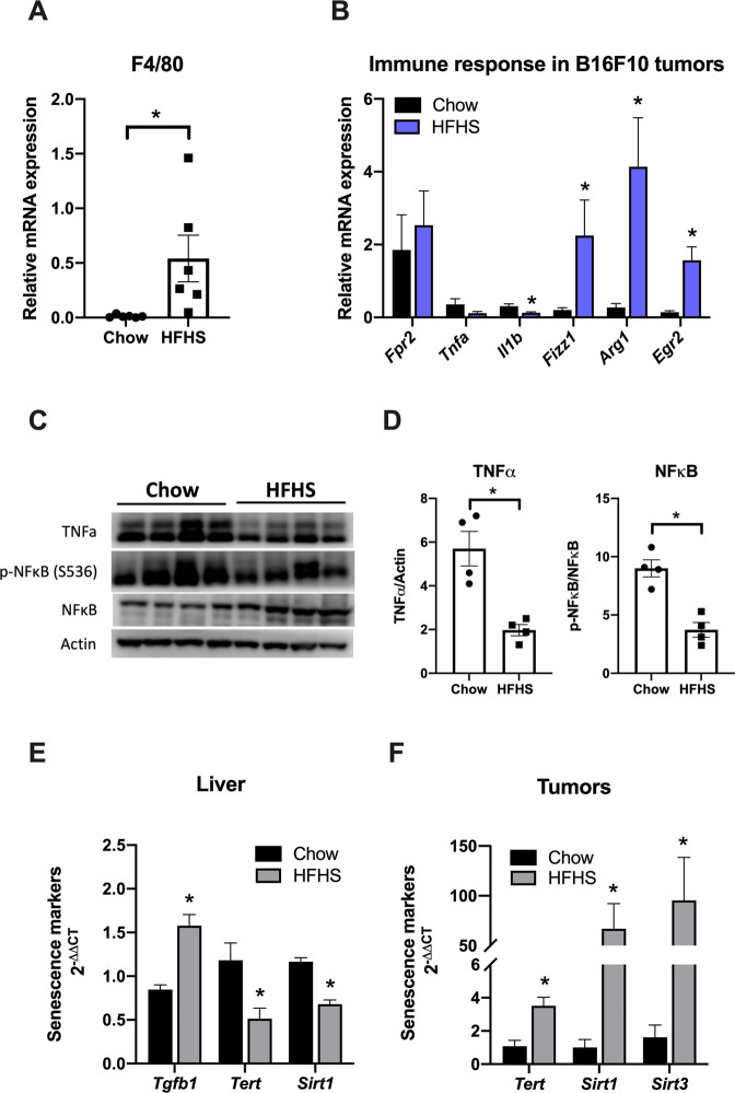
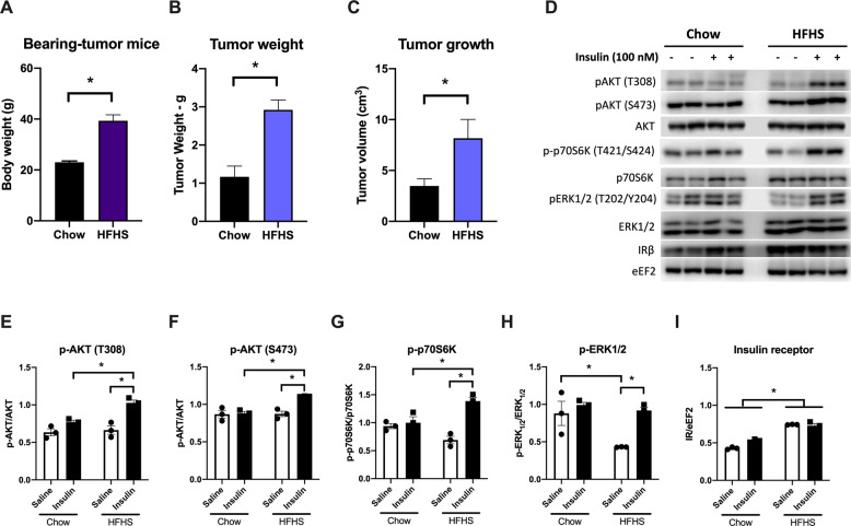
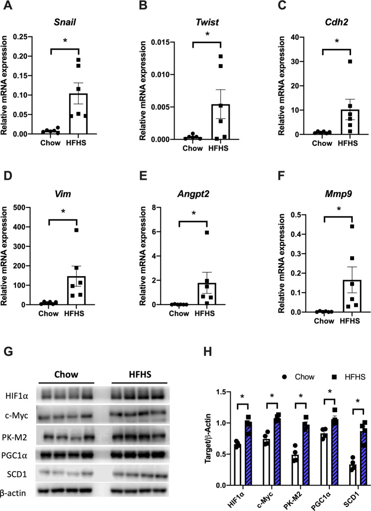
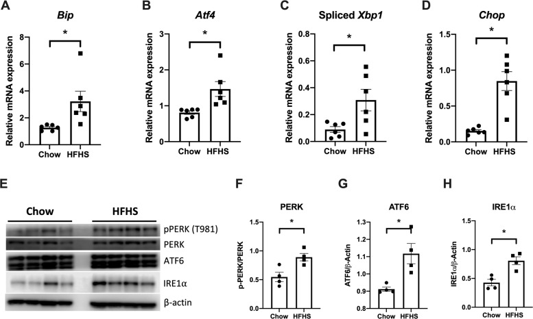
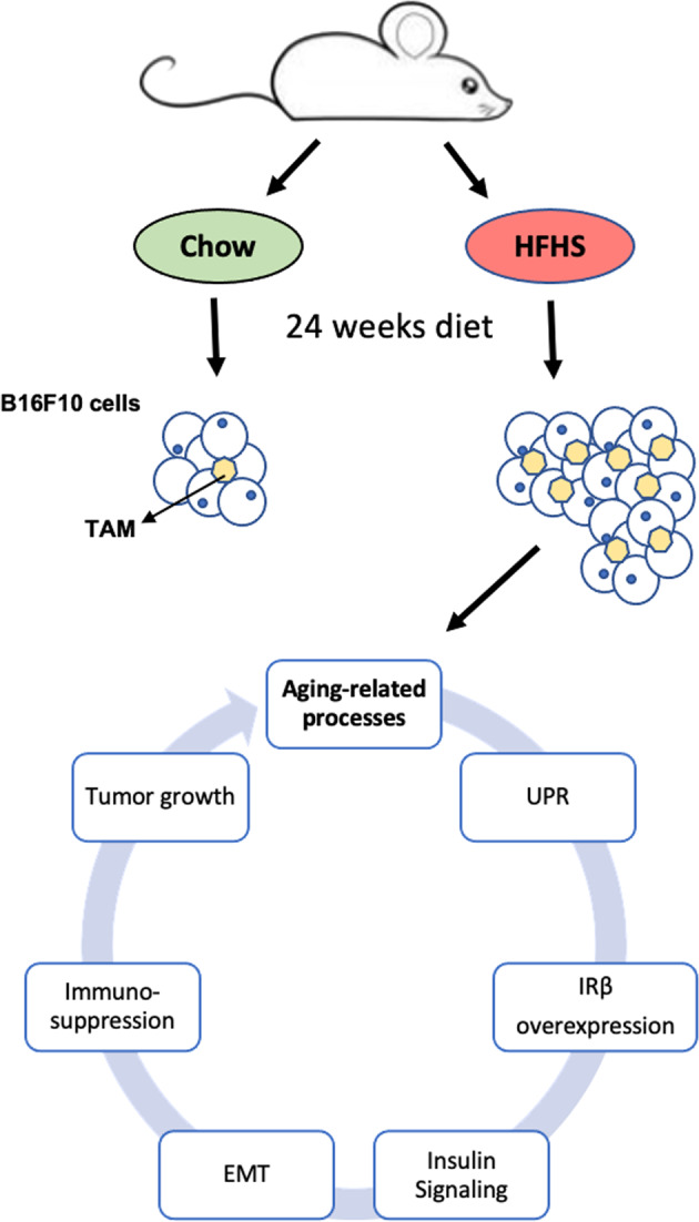
Among the principal causative factors for the development of complications related to aging is a diet rich in fats and sugars, also known as the Western diet. This diet advocates numerous changes that might increase the susceptibility to initiate cancer and/or to create a tissue microenvironment more conducive to the growth of malignant cells, thus favoring the progression of cancer and metastasis. Hypercaloric diets in general lead to oxidative stress generating reactive oxygen species and induce endoplasmic reticulum stress. Our results demonstrate that mice bearing tumors fed with a Western diet presented bigger tumor mass with increased insulin sensitivity in these tissues. Several markers of insulin signaling, such as AKT phosphorylation and mTOR pathway, are promoted in tumors of Western diet-fed animals. This process is associated with increased macrophage infiltration, activation of unfolded protein response pathway, and initiation of epithelial-mesenchymal transition (EMT) process in these tumor tissues. Summing up, we propose that the Western diet accelerates the aging-related processes favoring tumor development.


Conflict of interest statement

The authors declare no competing interests.

Figures

Fig. 1
Fig. 1. Macrophage infiltration, M2-like polarization, and anti-inflammatory response increase in tumors of HFHS-fed mice.
C57BL/6J mice were treated with chow or HFHS diet as described in “Materials and methods” section. Then, B16F10 cells (10,000 cells in 50 μL PBS) were implanted subcutaneously in the back of wild-type mice. After 3 weeks, mice were sacrificed and tumor tissues were subject to several analyses. mRNA levels of macrophage infiltration (F4/80 or Adgre1, A) and immune response (Fpr2, Tnfa, IL1b, Fizz1, Arg1, and Egr2, B) from tumor tissues were measured by qPCR analysis (n = 6). *P < 0.05 as compared to control (Mann–Whitney test). C Western blot analysis of proteins associated with tumor’ inflammatory response to HFHS diet (each lane in the Western blot represents a tumor from a different animal). D TNFα, pNF-κB (S536), and NF-κB were quantified using the ImageJ software and the quantification of each staining is plotted in the panels normalized by the quantification of the total protein staining. β-Actin was used as the loading control. Values are mean ± SEM of four independent animals (n = 4). *P < 0.05 as compared to control (unpaired Student’s t test). E Relative mRNA expression of Tgfb1, Tert, and Sirt1 from mice livers measured by qPCR analysis. F Relative mRNA expression of Tert, Sirt1, and Sirt3 from tumors measured by qPCR analysis (bars are mean 2−ΔΔCt ± SEM; n = 6; *P < 0.05, Mann–Whitney non-parametric test as compared to control).
Fig. 2
Fig. 2. HFHS diet significantly increases tumor weight and volume and mice body weight.
Twelve-week-old C57Bl6/6J female mice were fed standard chow or HFHS diet for 26 weeks. Then, B16F10 cells (10,000 cells in 50 μL PBS) were implanted subcutaneously in the back of wild-type mice. After 3 weeks, mice were killed and tumor tissues were subject to several analyses. A Body weight of the mice at the end of the experiment (g ± SEM). B Tumor weight (g ± SEM, n = 6). C Tumor volume measured with digital calipers (cm3 ± SEM). D Insulin signaling was analyzed by Western blots in protein lysates of the tumors from chow and HFHS-fed mice, which were treated with an insulin bolus after having food withheld for 6 h. Representative Western blot of total and phosphorylated AKT, p70S6K and ERK1/2, and insulin receptor β (IRβ) as shown for chow (left) and HFHS (right) group. EI pAKT (T308)/AKT, pAKT (S473)/AKT, p-p70S6K (T421/S424)/p70S6K, pERK1/2 (T202/Y204)/ERK1/2, and IRβ/eEF2 were quantified using the ImageJ software, and the quantification of each staining is plotted in the panels normalized by the quantification of the total protein staining. eEF2 was used as the loading control. Values are mean ± SEM of three independent animals (n = 3). *P < 0.05 as compared to control (unpaired Student’s t test for (AC), and two-way ANOVA, followed by Tukey’s post test for EI).
Fig. 3
Fig. 3. HFHS diet induces EMT, angiogenesis, and aggressiveness through oncogenic stimulation.
mRNA expression of EMT-related genes (Snail, Twist, Cdh2, and Vim, AD), angiogenesis (Angpt2, E), and matrix metalloproteinase 9 (Mmp9, F) were assessed by qPCR (n = 6). G Western blot of protein extract from tumor tissues of mice fed chow or HFHS diet. H Densitometric quantification of Western blots. Results are expressed as means ± SEM (n = 4). *P < 0.05 as compared to control (Mann–Whitney test for (AF) and unpaired Student’s t test for H).
Fig. 4
Fig. 4. ER stress and UPR activation by HFHS died in tumor tissues.
A–D mRNA expression of Bip, Atf4, spliced Xbp1, and Chop genes were assessed by qPCR. Results are expressed as means ± SEM (n = 6). E Western blot analysis of proteins associated with the UPR signaling pathway in tumors from chow or HFHS-fed mice. Representative Western blot of PERK, IRE1α, and ATF6 are shown for chow (left panel) and HFHS (right panel) groups. FH β-Actin was used as the loading control. Values are mean ± SEM of four independent animals (n = 4). All plotted values are statistically different from the chow group (Mann–Whitney test for panels AD and unpaired Student’s t test for panels FH).
Fig. 5
Fig. 5. Summary of the effects of HFHS diet on tumor biology and inflammation.
HFHS high-fat high-sucrose, TAM tumor-associated macrophage, UPR unfolded protein response, IRβ insulin receptor β, EMT epithelial–mesenchymal transition.

References

    1. Fane M, Weeraratna AT. How the ageing microenvironment influences tumour progression. Nat Rev Cancer. 2020;20:89–106. doi: 10.1038/s41568-019-0222-9. - DOI - PMC - PubMed
    1. Font-Burgada J, Sun B, Karin M. Obesity and cancer: the oil that feeds the flame. Cell Metab. 2016;23:48–62. doi: 10.1016/j.cmet.2015.12.015. - DOI - PubMed
    1. Dietz WH, Baur LA, Hall K, Puhl RM, Taveras EM, Uauy R, et al. Management of obesity: improvement of health-care training and systems for prevention and care. Lancet. 2015;385:2521–33. doi: 10.1016/S0140-6736(14)61748-7. - DOI - PubMed
    1. Laimer M, Melmer A, Lamina C, Raschenberger J, Adamovski P, Engl J, et al. Telomere length increase after weight loss induced by bariatric surgery: results from a 10 year prospective study. Int J Obes. 2016;40:773–8. doi: 10.1038/ijo.2015.238. - DOI - PubMed
    1. López-Otín C, Galluzzi L, Freije JMP, Madeo F, Kroemer G. Metabolic control of longevity. Cell. 2016;166:802–21. doi: 10.1016/j.cell.2016.07.031. - DOI - PubMed

Publication types

MeSH terms