. 2021 Jun 23;12(1):3902.
doi: 10.1038/s41467-021-24171-z.

Nucleation of protein mesocrystals via oriented attachment

Affiliations

Nucleation of protein mesocrystals via oriented attachment

Alexander E S Van Driessche et al. Nat Commun. .

Abstract

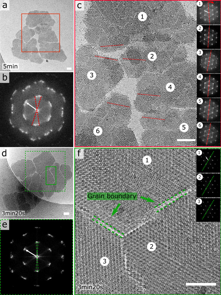
Self-assembly of proteins holds great promise for the bottom-up design and production of synthetic biomaterials. In conventional approaches, designer proteins are pre-programmed with specific recognition sites that drive the association process towards a desired organized state. Although proven effective, this approach poses restrictions on the complexity and material properties of the end-state. An alternative, hierarchical approach that has found wide adoption for inorganic systems, relies on the production of crystalline nanoparticles that become the building blocks of a next-level assembly process driven by oriented attachment (OA). As it stands, OA has not yet been observed for protein systems. Here we employ cryo-transmission electron microscopy (cryoEM) in the high nucleation rate limit of protein crystals and map the self-assembly route at molecular resolution. We observe the initial formation of facetted nanocrystals that merge lattices by means of OA alignment well before contact is made, satisfying non-trivial symmetry rules in the process. As these nanocrystalline assemblies grow larger we witness imperfect docking events leading to oriented aggregation into mesocrystalline assemblies. These observations highlight the underappreciated role of the interaction between crystalline nuclei, and the impact of OA on the crystallization process of proteins.


Conflict of interest statement

The authors declare no competing interests.

Figures

Fig. 1
Fig. 1. Oriented attachment of GI R387A nanocrystals.
a Submicron nanocrystals formed 1 min 40 s after mixing protein and precipitant; corresponding FFT images exhibit sharp maxima; b zoom-in of a particle rotated 90° with the c-axis in the plane of imaging (i.e. side view), showing 7 molecular rows with a lattice spacing along the c-axis of 8.4 nm; c oriented attachment of individually nucleated nanocrystals into a larger, merged lattice composed of domains 1 and 2, d making loose lateral contact with domain 3 at an angle of 4.3°; e two nanocrystals with merged lattices; red inset: FFT of the second molecular layer that is forming on the parent crystal; green inset: zoom-in of the unfinished molecular layer resolving local disorder and incoming growth units; f surface area of single (n = 60) and docked (n = 61) nanocrystals: center line, mean; box limits, upper and lower quartiles; whiskers, 1.5x interquartile range; points, outliers (g) and (h) large aligned nanocrystal assemblies with and without fault lines between the separate domain, respectively. Scalebar is 100 nm in panels a, b, c, d, e, g and h, and 50 nm in panel d. White arrows in the FFT’s correspond to a resolution of 6.6nm unless stated otherwise.
Fig. 2
Fig. 2. GI R387A mesocrystals.
a, c, d Large micron-sized composite nanocrystal structures with pronounced fault lines that separate lattice domains that are in near-alignment as demonstrated by their respective FFTs (b, e); zoom-in of the grain boundaries that span one or two molecular distances between the individual domains (f). The inset in panel (f) demonstrates the high degree of lattice order within each separate domain: domain 2 shown as representative example. Scalebar is 50 nm in panels a, c and d, and 25 nm in panel (f). White arrows in the FFT’s correspond to a resolution of 6.6 nm.
Fig. 3
Fig. 3. Lack of inter-crystal alignment in large nanocrystal assemblies.
a Poly-crystalline cluster with local hotspots of alignment (see FFT insets), b example of two vertically stacked domains exhibiting registry between their corresponding lattices (blue) but a 26° misalignment with the region enclosed in red, c example of lack of axial alignment between two nanocrystals where the FFT of the interlaced pattern (green) reveals two independent lattices residing at an angle of 25°, d disordered grouping of over 100 nanocrystals. Scalebar is 100 nm. White arrows in the FFT’s correspond to a resolution of 6.6 nm.
Fig. 4
Fig. 4. Model for OA of GI nanocrystals.
GI molecules pack along a 3-fold axis within the (001) plane in which we discern three different GI orientations (1,2,3). Nearest neighbors exclude GI molecules with identical orientations; a Simplified scheme of self-assembly: freely diffusing nanocrystals approach each other, followed by rotational and translational adjustments to align both lattices. Alignment facilitates a final jump to contact by desolvation of the surface patches that partake in lattice contact formation; b Illustration of three different scenarios for further growth: I and II violate H32 symmetry rules and are likely to lead to the formation of a GB at the interface; III leads to successful merger of all three lattices and the resulting voids can be filled by monomer addition.
Fig. 5
Fig. 5. Protein crystal nucleation pathways that have been experimentally observed at the nanoscale.
P1: one-step nucleation solely involving crystalline clusters throughout the entire pathway (glucose isomerase I222); P2 and P3: involving oriented attachment of 2D, 3D and 1D crystalline clusters into larger ordered assemblies (glucose isomerase H32 and P2 2 2); P3*: spinodal decomposition limit of the P3 scenario leading to kinetic jamming (gel); P4: self-seeded nucleation of crystalline clusters on the surfaces of solid, amorphous condensates (lysozyme); P5: two-step nucleation comprising initial densification into loose disordered clusters, followed by gradual local desolvation and densification into a crystalline array (ferritin); P6: aggregation in the high supersaturation limit with poorly ordered clusters.

References

    1. Kashchiev, D. Nucleation - Basic Theory with Applications (Butterworth-Heinemann, 2000).
    1. Karthika S, Radhakrishnan TK, Kalaichelvi P. A review of classical and nonclassical nucleation theories. Cryst. Growth Des. 2016;16:6663–6681. doi: 10.1021/acs.cgd.6b00794. - DOI
    1. Van Driessche, A., Kellermeier, M., Benning, L. G., Gebauer, D. New Perspectives on Mineral Nucleation and Growth: From Solution Precursors to Solid Materials (Springer International Publishing, 2017).
    1. Chung S, Shin S-H, Bertozzi CR, Yoreo JJD. Self-catalyzed growth of S layers via an amorphous-to-crystalline transition limited by folding kinetics. Proc. Natl Acad. Sci. USA. 2010;107:16536–16541. doi: 10.1073/pnas.1008280107. - DOI - PMC - PubMed
    1. Van Driessche AES, et al. Molecular nucleation mechanisms and control strategies for crystal polymorph selection. Nature. 2018;556:89–94. doi: 10.1038/nature25971. - DOI - PubMed

Publication types