. 2021 Jul;595(7867):404-408.
doi: 10.1038/s41586-021-03672-3. Epub 2021 Jun 23.

Mechanism of disease and therapeutic rescue of Dok7 congenital myasthenia

Affiliations

Mechanism of disease and therapeutic rescue of Dok7 congenital myasthenia

Julien Oury et al. Nature. 2021 Jul.

Abstract

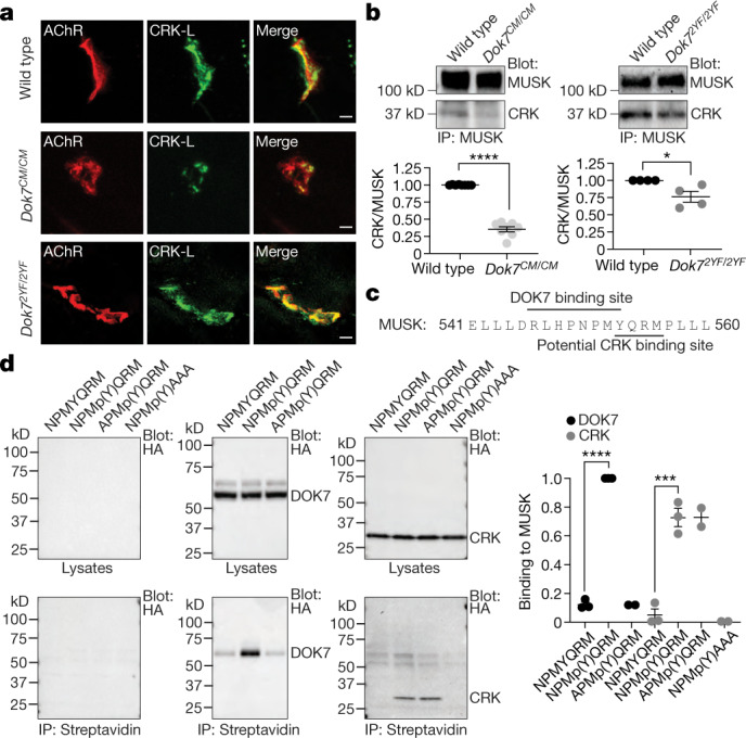
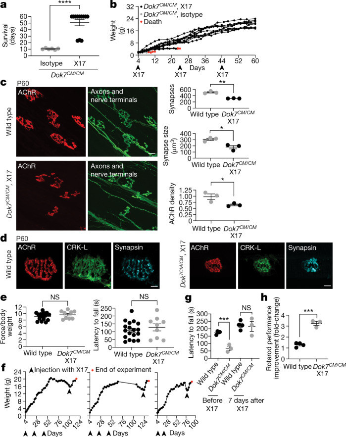
Congenital myasthenia (CM) is a devastating neuromuscular disease, and mutations in DOK7, an adaptor protein that is crucial for forming and maintaining neuromuscular synapses, are a major cause of CM1,2. The most common disease-causing mutation (DOK71124_1127 dup) truncates DOK7 and leads to the loss of two tyrosine residues that are phosphorylated and recruit CRK proteins, which are important for anchoring acetylcholine receptors at synapses. Here we describe a mouse model of this common form of CM (Dok7CM mice) and a mouse with point mutations in the two tyrosine residues (Dok72YF). We show that Dok7CM mice had severe deficits in neuromuscular synapse formation that caused neonatal lethality. Unexpectedly, these deficits were due to a severe deficiency in phosphorylation and activation of muscle-specific kinase (MUSK) rather than a deficiency in DOK7 tyrosine phosphorylation. We developed agonist antibodies against MUSK and show that these antibodies restored neuromuscular synapse formation and prevented neonatal lethality and late-onset disease in Dok7CM mice. These findings identify an unexpected cause for disease and a potential therapy for both DOK7 CM and other forms of CM caused by mutations in AGRIN, LRP4 or MUSK, and illustrate the potential of targeted therapy to rescue congenital lethality.


Conflict of interest statement

S.J.B. is an inventor on a patent (no. 9,329,182) for ‘Method of treating motor neuron disease with an antibody that agonizes MUSK’. S.J.B., S.K., J.O., A.K. and N.L. are inventors on a patent application (no. 1474662.02232) for ‘Therapeutic MUSK Antibodies’ filed by New York University. These patents have been licensed to Argenx BVBA. S.J.B. and S.K. received research funding from Argenx BVBA. S.K. is a scientific advisory board member and holds equity in and receives consulting fees from Black Diamond Therapeutics, and receives research funding from Puretech Health.

Figures

Fig. 1
Fig. 1. The C-terminal region of DOK7 is essential for synaptic differentiation and to sustain MUSK tyrosine phosphorylation.
a, Dok71124_1127,dup (Dok7CM) leads to a frame-shift and premature termination, including loss of Y396 and Y406. b, Expected and observed numbers of progeny, and χ2 values, from intercrossing Dok7 heterozygous mutant mice. c, d, Staining of AChRs (red) and axons and nerve terminals (green) in diaphragm muscles from wild-type and Dok7 mutant E18.5 mice (c). Scale bars, 10 μm. Scatter plots (d) show the number of synapses, synaptic size, and density of synaptic AChRs from n = 5–8 mice of each genotype. e, f, Western blot (top) and quantification (bottom) of DOK7 expression in E18.5 mice. n = 8 mice in e, 11 in f. t-DOK7, truncated DOK7; IP, immunoprecipitation. g, h, Western blot (top) and quantification (bottom) of MUSK tyrosine phosphorylation (pTyr) in E18.5 mice. n = 7 mice in g, 5 in h. Plots show data for individual mice and mean ± s.e.m.; NS, not significant; ****P < 0.00005; two-sided Student’s t-test. Source data
Fig. 2
Fig. 2. Recruitment of CRK to the synapse and to the MUSK–DOK7 complex is impaired in Dok7CM/CM mice.
a, Staining for CRK-L (green) and AChRs (red) in muscle sections from E18.5 mice. Scale bars, 5 μm. Representative images from three experiments. b, Top, co-immunoprecipitation of CRK with MUSK from muscles of E18.5 mice. Bottom, CRK levels normalized to MUSK levels for mice of each genotype (n = 8 for Dok7CM/CM and n = 4 for Dok72YF/2YFmice). c, Amino acid sequence of the MUSK JM region showing binding site for DOK7 and potential binding site for CRK. d, Left, affinity capture of DOK7 and CRK-I with phosphorylated and non-phosphorylated peptides detected by immunoblotting. The peptide sequences are shown. Right, quantification. Plots show individual data and mean ± s.e.m.; *P < 0.05, ***P < 0.0005, ****P < 0.00005; two-sided Student’s t-test. Source data
Fig. 3
Fig. 3. Antibodies against MUSK Fz domain stimulate MUSK phosphorylation in cultured myotubes and bind MUSK in vivo.
a, MUSK phosphorylation, normalized to MUSK expression, in C2 myotubes treated for 30 min with biotinylated Fabs each tetramerized with streptavidin. Top, western blot; bottom, quantification. b, MUSK phosphorylation in C2 myotubes treated with 0.5 nM AGRIN or 10 nM antibodies, with either mouse IgG2a or human IgG1 Fc regions. Top, western blot; bottom, quantification. c, Left, staining for AChRs (red) and human IgG (cyan) in diaphragm muscles of P30 wild-type mice two days after intraperitoneal injection of antibody X17. Right, X17 signal intensities normalized to AChR plotted against antibody dose (n = 3 mice per concentration). Plots show mean ± s.e.m. (with individual data points in a, b). Source data
Fig. 4
Fig. 4. An agonist antibody against MUSK restores synapse development and rescues lethality in young Dok7CM mice and reverses disease relapse in adult Dok7CM mice.
a, Dok7CM/CM mice on a C57BL/6-CBA mixed background (n = 12) were injected with X17 at P4, P24 and P44, and the experiment was ended when the mice were at P60. Mice (n = 6) injected with the isotype control died within two weeks of birth. b, X17 restored weight gain in Dok7CM/CM mice. c, Left, diaphragm muscles from P60 mice stained for AChRs (red) and neurofilament and synapsin to label motor axons and nerve terminals (green). Scale bar, 10 μm. Right, quantification (n = 3 mice, >50 synapses per mouse). d, Staining for AChRs (red), CRK-L (green) and synapsin (cyan) in myofibres isolated from muscles of wild-type mice (left) and Dok7CM/CM mice injected with X17 (right). Representative images of ten synapses per mouse from three mice. Scale bars, 5 μm. e, Grip strength and latency to fall from a rotating rotarod of Dok7CM/CM mice treated with X17 (n = 9), compared with wild-type mice (n = 18). f, Weight changes in Dok7CM/CM mice treated either with mIgG2a-X17 at P4, P24 and P44 or with hIgG1-X17 at P4 and P18, with antibody treatment then discontinued for 2–3 months. When Dok7CM/CM mice began to lose weight and showed motor deficits, they were re-injected with hIgG1-X17 and their weights monitored. Red dots indicate death at the end of the experiment. g, Latency to fall for four wild-type and three Dok7CM/CM mice. h, Change in rotarod performance one week after X17 treatment of Dok7CM/CM mice (fold-change from performance before X17), compared with that of wild-type mice not injected with X17. All plots except b, f show individual data points and mean ± s.e.m.; NS, not significant; *P < 0.05, **P < 0.005, ***P < 0.0005, ****P < 0.00005; two-sided Student’s t-test. Source data
Extended Data Fig. 1
Extended Data Fig. 1. Characterization of neuromuscular synapses in Dok7 mutant mice.
a, b, Left, diaphragm muscles from E18.5 (a) and P135 (b) wild-type and Dok7 mutant mice were stained with Alexa 488–anti-BGT to label AChRs (red) and antibodies against neurofilament and synapsin to label motor axons and nerve terminals (green). Scale bars, 50 μm (a), 10 μm (b). a, Right, endplate width, denervation and co-localization of synapses in wild-type, Dok7CM/CM and Dok72YF/2YF mice. The width of the endplate band (dashed lines) was increased by 45% in Dok7CM/CM mice but was normal in Dok72YF/2YF mice. In Dok7CM/CM mice, 17% of AChR clusters were completely unopposed by nerve terminals, indicating denervated myofibres. Many synapses in Dok7CM/CM mice were partially innervated, as nearly half of the AChR-rich area at synapses was not juxtaposed by nerve terminals. b, Right, in Dok72YF/2YF mice, synapses mature from a plaque-like to a complex, pretzel-like shape, characteristic of mature mouse neuromuscular synapses. Synapses in Dok72YF/2YF mice, however, often appeared elongated. The number of synapses and the density of synaptic AChRs were similar in wild-type and Dok72YF/2YF mice. Synaptic size was increased by 20% in Dok72YF/2YF mice when compared with wild-type mice. Data shown as mean ± s.e.m. from 3 mice (>50 synapses per mouse). n.s., not significant; *P < 0.05, ****P < 0.00005; two-sided Student’s t-test. Source data
Extended Data Fig. 2
Extended Data Fig. 2. Wild-type and truncated DOK7 are detected with similar efficiency by antibodies against the PH and PTB domains in DOK7.
a, HEK 293 cells were transiently transfected with a plasmid expressing either HA-tagged DOK7 or HA-tagged truncated DOK7 encoded by Dok71124_1127 TGCC dup. Proteins in cell lysates (triplicates) were separated by SDS–PAGE, and western blots were probed with either a rabbit antibody against the PTB domain in DOK7 or a monoclonal antibody against HA (left). We measured the grey levels of the bands for wild-type and truncated DOK7 proteins and normalized the level detected by western blotting with the rabbit antibody against DOK7 with the level detected by western blotting with the antibody against HA. The ratio for wild-type DOK7 was equivalent to the ratio for truncated DOK7 (left), indicating that the rabbit antibody against DOK7 detected wild-type and truncated DOK7 proteins with similar efficiency by western blotting. b, Wild-type and truncated DOK7 were immunoprecipitated with similar efficiency by a goat antibody against the PTB domain in DOK7. HEK 293 cells were transiently co-transfected with plasmids expressing HA-tagged DOK7 and HA-tagged truncated DOK7 encoded by Dok71124_1127 TGCC dup. DOK7 proteins were immunoprecipitated from cell lysates (triplicates) with either a monoclonal antibody against HA or a goat antibody against the PTB domain in DOK7, and western blots were probed with the monoclonal antibody against HA (left). We measured the grey levels, subtracted the level for the background band in the control, non-transfected samples, and normalized the value for each protein immunoprecipitated with the goat-antibody against DOK7 to the value for the same protein immunoprecipitated with the antibody against HA. This ratio was equivalent for wild-type and truncated DOK7 proteins, indicating that the goat antibody against DOK7 immunoprecipitated wild-type and truncated proteins with similar efficiency (right). Plots show individual values from three and four experiments for a and b, respectively, and the mean ± s.e.m. (n.s., not significant); two-sided Student’s t-test. Source data
Extended Data Fig. 3
Extended Data Fig. 3. Dok7 RNA expression is normal in Dok7CM/CM mice.
a, RT–PCR amplification of Dok7 RNA shows that Dok7 mRNA levels are similar in muscle from E18.5 wild-type and Dok7CM/CM mice. GAPDH was used as a loading control. b, Dok7 mRNA levels were quantified by qPCR, which showed that Dok7 mRNA levels are normal in Dok7CM/CM mice (n = 3 mice). c, DOK7 was immunoprecipitated from muscles of E18.5 wild-type and Dok7CM/CM mice, and the blots were probed with antibodies against DOK7 (left). Truncated DOK7, encoded by Dok7CM/CM, migrates at the predicted size, but is expressed at threefold lower levels than wild-type DOK7 (right; n = 10 mice). Data shown as individual data points and mean ± s.e.m.; n.s., not significant; ****P < 0.00005; two-sided Student’s t-test. Source data
Extended Data Fig. 4
Extended Data Fig. 4. Y396 and Y406 are the main, if not sole, tyrosine residues in DOK7 that are phosphorylated by AGRIN stimulation.
We generated muscle cell lines from wild-type and Dok72YF/2YF mice and treated the cultured myotubes with AGRIN for 30 min. MUSK was immunoprecipitated, and western blots were probed with antibodies against MUSK or phosphotyrosine (pTyr). AGRIN stimulates DOK7 tyrosine phosphorylation in wild-type but not Dok72YF/2YF myotubes (data from two experiments).
Extended Data Fig. 5
Extended Data Fig. 5. MUSK antibody clones.
a, Amino acid sequences of the complementarity-determining regions (CDRs) of antibodies against MUSK developed in this study. CDR definitions are based on Wu and Kabat, except that CDR-H1 includes four additional residues at the N terminus to show diversified positions. b, Amino acid sequences of the VL and VH domains of clone X17. c, d, Binding titration of antibodies against MUSK in the Fab format to immobilized hFz, hECD, mFz and mECD, as tested using a bead-based binding assay. Curves show the best fit of the 1:1 binding model. The table lists apparent KD values (mean ± s.d., n = 3). The datasets in c and d were obtained different instruments, which resulted in different signal ranges. e, Binding titration of antibodies against MUSK in the IgG format, as in c. Source data
Extended Data Fig. 6
Extended Data Fig. 6. Chronic injection of MUSK agonist antibody X17 in wild-type mice has no effect on the organization of neuromuscular synapses, weight gain or motor behaviour.
a, Blood half-life measurements of X17-mIgG2a-LALAPG. Nonlinear least-squares fitting of the median fluorescence intensities with a single exponential curve for three mice is shown. The half-life was determined to be 4.9 ± 0.2 days. b, Wild-type mice on a C57BL/6-CBA mixed background, injected at P4, P24 and P44 with X17 (n = 4), survived until P60, when the experiment was ended. The scatter plot shows the survival time for nine non-injected wild-type mice and four wild-type mice injected with X17 with mean ± s.e.m. (n.s., not significant). c, Wild-type mice injected with X17 (n = 4) gained weight like uninjected wild-type mice (n = 9). d, Left, diaphragm muscles from P60 wild-type mice and wild-type mice injected with X17 were stained with Alexa 488–anti-BGT to label AChRs (red) and antibodies against neurofilament and synapsin to label motor axons and nerve terminals (green). In wild-type mice treated with X17, synapses matured from a simple, plaque-like shape to a complex, pretzel-like shape, characteristic of mature mouse neuromuscular synapses. Scale bar, 10 μm. Right, injection of X17 in wild-type mice had no effect on synapse number, size or AChR density. We analysed more than 50 synapses per diaphragm muscle from two mice in each category. e, Motor performance of wild-type mice injected with X17, as assessed by grip strength and the latency to fall from a rotating rotarod, was similar to that of non-injected wild-type mice. The scatter plots show the values for 18 wild-type mice and 4 wild-type mice injected with X17 and the mean ± s.e.m; two-sided Student’s t-test. Source data
Extended Data Fig. 7
Extended Data Fig. 7. The C-terminal region of DOK7 is essential for complete differentiation and maturation of the neuromuscular synapse in Dok7CM/CM mice on a mixed genetic background.
ac, Left, diaphragm muscles from wild-type and Dok7CM/CM mice on a C57BL/6-CBA mixed background at E18.5 and P10 were stained with Alexa 488–anti-BGT to label AChRs (red) and antibodies against neurofilament and synapsin to label motor axons and nerve terminals (green). Scale bars, 50 μm (a), 10 μm (b, c). a, Right, at E18.5, the endplate band (dashed white lines on left) is 30% wider in Dok7CM/CM than wild-type mice. Moreover, nerve terminals were absent from 15% of AChR clusters and the colocalization index (synapsin/AChR) was reduced 3.5-fold in Dok7CM/CM mice. n = 3 mice. b, Right, the number of synapses, synaptic size and density of synaptic AChRs were reduced 3.2-, 4.5- and 8-fold, respectively, in E18.5 Dok7CM/CM mice. n = 3 mice (>50 synapses per mouse). c, Right, at P10, the number of synapses, synaptic size and density of synaptic AChRs were reduced over tenfold in Dok7CM/CM mice. In addition, nerve terminals were absent from 20% of the AChR clusters in Dok7CM/CM mice. n = 3 mice (>50 synapses per mouse). d, DOK7 was immunoprecipitated from muscles of E18.5 wild-type and Dok7CM/CM mice, and the blots were probed with antibodies against DOK7 (left). Truncated DOK7, encoded by Dok7CM/CM, migrated at the predicted size, but was expressed at threefold lower levels than wild-type DOK7 (right). Because DOK7 expression and MUSK phosphorylation were diminished to the same extent in the C57BL/6-CBA mixed breed and C57BL/6 inbred mice, other factors presumably led to increased survival in the mixed genetic background. n = 8 mice per genotype. e, MUSK was immunoprecipitated from muscles of E18.5 wild-type and Dok7CM/CM mice, and the blots were probed with antibodies against MUSK, phosphotyrosine, and CRK (left). The levels of phosphotyrosine and CRK that co-isolated with the MUSK complex were normalized to MUSK expression (right). CRK association with the MUSK complex was 2.8-fold lower in Dok7CM/CM mice than wild-type mice. MUSK tyrosine phosphorylation was fivefold lower in Dok7CM/CM mice than wild-type mice. n = 3 mice per genotype. Scatter plots show individual data points and mean ± s.e.m.; *P < 0.05, **P < 0.005, ***P < 0.0005, ****P < 0.00005; two-sided Student’s t-test. Source data
Extended Data Fig. 8
Extended Data Fig. 8. Antibodies X2 and X3, like X17, rescue Dok7CM/CM mice from early lethality.
a, Dok7CM/CM mice on a C57BL/6-CBA mixed background were injected at P4 with 10 mg kg−1 mIgG2a-X3. At this dose, X3 failed to rescue the mice from lethality. b, By contrast, dosing with 20 mg kg−1 mIgG2a-X3 at P4 rescued the mice from early lethality. These mice were subsequently injected with 10 mg kg−1 mIgG2a-X3 at P18, which led to survival until P60, when the experiment was ended. c, Injecting Dok7CM/CM mice with 20 mg kg−1 hIgG1-X2 at P4 likewise rescued Dok7CM/CM mice from early lethality; subsequent injection of 10 mg kg−1 hIgG1-X2 at P18 led to survival of Dok7CM/CM mice until P60, when the experiment was ended. Source data

Comment in

References

    1. Müller, J. S., Mihaylova, V., Abicht, A. & Lochmüller, H. Congenital myasthenic syndromes: spotlight on genetic defects of neuromuscular transmission. Expert Rev. Mol. Med. 9, 1–20 (2007).10.1017/S1462399407000427 - DOI - PubMed
    1. Engel, A. G., Shen, X. M., Selcen, D. & Sine, S. M. Congenital myasthenic syndromes: pathogenesis, diagnosis, and treatment. Lancet Neurol. 14, 420–434 (2015). 10.1016/S1474-4422(14)70201-7 - DOI - PMC - PubMed
    1. Sanes, J. R. & Lichtman, J. W. Induction, assembly, maturation and maintenance of a postsynaptic apparatus. Nat. Rev. Neurosci. 2, 791–805 (2001). 10.1038/35097557 - DOI - PubMed
    1. Burden, S. J., Yumoto, N. & Zhang, W. The role of MuSK in synapse formation and neuromuscular disease. Cold Spring Harb. Perspect. Biol. 5, a009167 (2013). 10.1101/cshperspect.a009167 - DOI - PMC - PubMed
    1. Tintignac, L. A., Brenner, H. R. & Rüegg, M. A. Mechanisms regulating neuromuscular junction development and function and causes of muscle wasting. Physiol. Rev. 95, 809–852 (2015). 10.1152/physrev.00033.2014 - DOI - PubMed

Publication types

MeSH terms