. 2021 Jun 7:12:683902.
doi: 10.3389/fimmu.2021.683902. eCollection 2021.

Hyper-Enriched Anti-RSV Immunoglobulins Nasally Administered: A Promising Approach for Respiratory Syncytial Virus Prophylaxis

Affiliations

Hyper-Enriched Anti-RSV Immunoglobulins Nasally Administered: A Promising Approach for Respiratory Syncytial Virus Prophylaxis

Emilie Jacque et al. Front Immunol. .

Abstract

Respiratory syncytial virus (RSV) is a public health concern that causes acute lower respiratory tract infection. So far, no vaccine candidate under development has reached the market and the only licensed product to prevent RSV infection in at-risk infants and young children is a monoclonal antibody (Synagis®). Polyclonal human anti-RSV hyper-immune immunoglobulins (Igs) have also been used but were superseded by Synagis® owing to their low titer and large infused volume. Here we report a new drug class of immunoglobulins, derived from human non hyper-immune plasma that was generated by an innovative bioprocess, called Ig cracking, combining expertises in plasma-derived products and affinity chromatography. By using the RSV fusion protein (F protein) as ligand, the Ig cracking process provided a purified and concentrated product, designated hyper-enriched anti-RSV IgG, composed of at least 15-20% target-specific-antibodies from normal plasma. These anti-RSV Ig displayed a strong in vitro neutralization effect on RSV replication. Moreover, we described a novel prophylactic strategy based on local nasal administration of this unique hyper-enriched anti-RSV IgG solution using a mouse model of infection with bioluminescent RSV. Our results demonstrated that very low doses of hyper-enriched anti-RSV IgG can be administered locally to ensure rapid and efficient inhibition of virus infection. Thus, the general hyper-enriched Ig concept appeared a promising approach and might provide solutions to prevent and treat other infectious diseases.

Importance: Respiratory Syncytial Virus (RSV) is the major cause of acute lower respiratory infections in children, and is also recognized as a cause of morbidity in the elderly. There are still no vaccines and no efficient antiviral therapy against this virus. Here, we described an approach of passive immunization with a new class of hyper-enriched anti-RSV immunoglobulins (Ig) manufactured from human normal plasma. This new class of immunoglobulin plasma derived product is generated by an innovative bioprocess, called Ig cracking, which requires a combination of expertise in both plasma derived products and affinity chromatography. The strong efficacy in a small volume of these hyper-enriched anti-RSV IgG to inhibit the viral infection was demonstrated using a mouse model. This new class of immunoglobulin plasma-derived products could be applied to other pathogens to address specific therapeutic needs in the field of infectious diseases or even pandemics, such as COVID-19.

Keywords: bioprocess; concentration; human plasma; hyper-immune immunoglobulins; nasal administration; neutralization; prophylactic strategy; viral infection.


Conflict of interest statement

The authors declare that the research was conducted in the absence of any commercial or financial relationships that could be construed as a potential conflict of interest.

Figures

Figure 1
Figure 1
Hyper-enriched anti-RSV Ig manufacturing. (A) General approach for anti-RSV Ig manufacturing. (B) Principle of the affinity step. (C) Hyper-enriched anti-RSV Ig affinity step chromatogram. This figure illustrates the reproducibility of the affinity step with the chromatogram superposition of two independent batches. (D) The integrity of the hyper-enriched anti-RSV Ig was compared to a commercial normal polyclonal IgG (ClariYg ®) by Coomassie blue staining SDS-PAGE.
Figure 2
Figure 2
Hyper-enriched anti-RSV Ig characterization. (A, B) The biological activity of anti-RSV Ig products was measured by in vitro neutralization test assessed on HEp-2 cells and using recombinant RSV-cherry. (A) The antiviral activity of the hyper-enriched anti-RSV Ig solution was compared to the starting solution (IVIg) and the antibody control, Synagis® . (B) The neutralizing capacity of two batches of hyper-enriched anti-RSV Ig was compared to Synagis®. Data are representative of 2 independent assays. (C) To determine the ability of the anti-RSV Ig product to neutralize a clinical isolate of RSV, the neutralization assay was performed by flow cytometry on HEp-2 infected cells after immunodetection of RSV-F protein. Results were expressed in MFI, 100 arbitrarily being the MFI obtained with RSV-infected HEp-2 cells without incubation with anti-RSV antibodies. Data are representative of 2 independent assays.
Figure 3
Figure 3
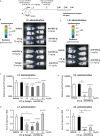
Evaluation of the protective efficacy of anti-RSV Ig in mouse infection model. (A) Experimental design showing the time and mode of RSV Ig delivery as well as the time of infection with RSV-Luc and the follow up of virus replication. (B–D) BALB/c mice received I.P. administration of 0.1, 1 or 10 mg/kg of anti-RSV Ig or 10 mg/kg of Synagis® before intranasal infection with RSV-luciferase (7 x 10⁵ pfu, 50 µL). As a control of protection/infection, mice were respectively treated with CTL Ig (10 mg/kg) or vehicle. (E–G) BALB/c mice received I.N. administration of 0.1 or 1 mg/kg of anti-RSV Ig (50 µL) before intranasal infection with RSV-luciferase (7 x 10⁵ pfu, 50 µL). As a control of protection/infection, mice were respectively treated with CTL Ig (1 mg/kg) or vehicle. (B, E) In vivo bioluminescence intensity was evaluated at 3, 4 and 5 d.p.i. with IVIS imaging system and is shown at 4 d.p.i. in this figure (dorsal view detection). Depicted images are representative of 2 independent pictures. (C, F) In vitro luciferase activity was measured in lung homogenates by quantification of bioluminescence emission (radiance in photon/sec/cm2/sr) using “Living Image” software after addition of D-luciferin to the lysates, and was normalized to the organ weight. Data are mean ± SEM from n = 4 mice. (D, G) mRNA level of RSV N gene was evaluated by real time RT-PCR in the lungs. Data are mean ± SEM from n = 4 mice. Significance is represented on graphs: *P < 0.05.
Figure 4
Figure 4
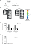
The RSV infection model of the upper respiratory tract. (A) BALB/c mice received intranasally a small inoculum volume (10 µL) of RSV-Luc (1.4 × 10⁵ pfu/mice) or mock (supernatant of Hep2 cells) to restrain viral replication to the upper respiratory tract. (B) Bioluminescence was measured at 1 and 4 d.p.i. by intranasal injection of 50 μl of D-luciferin and the capture of photon emission (dorsal view) from the whole animal using the IVIS system. (C) Luciferase activity was measured in NT or lungs homogenates by quantification of bioluminescence emission (radiance in photon/sec/cm2/sr) using “Living Image” software after addition of D-luciferin in the lysates. (D) mRNA level of RSV N gene was evaluated by real time RT-PCR in the lungs and expressed with the formula (2-ΔCt). Data are mean ± SEM from n = 3 mock-infected mice or n = 5 RSV-Luc-infected mice.
Figure 5
Figure 5
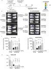
Efficacy of anti-RSV Ig intranasal administration at low doses on upper respiratory tract RSV infection in mice. (A) BALB/c mice received I.N. administration of 0.01 to 1 mg/kg of anti-RSV Ig in 10 µL one hour before I.N. injection of RSV-luciferase (1.4 x 10⁵ pfu, 10 µL). As a control of RSV-Ig, mice were treated with CTL Ig (0,1 or 1 mg/kg). (B) In vivo bioluminescence intensity was evaluated at 4 d.p.i. with IVIS imaging system (dorsal view detection). Depicted images are representative of 3 independent pictures. (C) Luciferase activity was measured in NT and lung homogenates by quantification of bioluminescence emission (radiance in photon/sec/cm2/sr) using “Living Image” software after addition of luciferin in the lysates, and was normalized to the organ weight. (D) mRNA level of RSV N gene was evaluated by real time RT-PCR in the lungs and expressed with the formula (2-ΔCt). Data are mean ± SEM from n = 6 mice for CTL Ig-treated mice or all doses of anti-RSV Ig and n = 12 for mice treated with 0.1 mg/kg of anti-RSV Ig. Significance is represented on graphs: *P < 0.05; **P < 0.01; ***P < 0.001.
Figure 6
Figure 6
Preventive effect of anti-RSV Ig intranasal administration on upper respiratory tract RSV infection in mice. (A) BALB/c mice received I.N. administration of 0.025 mg/kg of anti-RSV Ig in 10 µL five hours before I.N. injection of RSV-luciferase (1.4 x 10⁵ pfu, 10 µL). As a control of infection, mice were treated with CTL Ig. (B) In vivo bioluminescence intensity was evaluated at 1 d.p.i. with IVIS imaging system (dorsal view detection). (C) Luciferase activity was measured in NT and lung homogenates by quantification of bioluminescence emission (radiance in photon/sec/cm2/sr) using “Living Image” software after addition of luciferin in the wells, and was normalized to the organ weight. Data are mean ± SEM from n = 6 mice for CTL Ig-treated mice and n = 5 for mice treated with 0.025 mg/kg of anti-RSV Ig. Significance is represented on graphs: **P < 0.01.

References

    1. Vitiello G, Emmi G, Silvestri E, Di Scala G, Palterer B, Parronchi P. Intravenous Immunoglobulin Therapy: A Snapshot for the Internist. Intern Emerg Med (2019) 14:1041–9. 10.1007/s11739-019-02150-z - DOI - PubMed
    1. Eijkhout HW, van Der Meer JW, Kallenberg CG, Weening RS, van Dissel JT, Sanders LA, et al. . Inter-University Working Party for the Study of Immune: The Effect of Two Different Dosages of Intravenous Immunoglobulin on the Incidence of Recurrent Infections in Patients With Primary Hypogammaglobulinemia. A Randomized, Double-Blind, Multicenter Crossover Trial. Ann Intern Med (2001) 135(3):165–74. 10.7326/0003-4819-135-3-200108070-00008 - DOI - PubMed
    1. Bozzo J, Jorquera JI. Use of Human Immunoglobulins as an Anti-Infective Treatment: The Experience So Far and Their Possible Re-Emerging Role. Expert Rev Anti Infect Ther (2017) 15(6):585–604. 10.1080/14787210.2017.1328278 - DOI - PubMed
    1. Pelfrene E, Mura M, Cavaleiro Sanches A, Cavaleri M. Monoclonal Antibodies as Anti-Infective Products: A Promising Future? Clin Microbiol Infect (2019) 25(1):60–4. 10.1016/j.cmi.2018.04.024 - DOI - PMC - PubMed
    1. Zhu Q, McAuliffe JM, Patel NK, Palmer-Hill FJ, Yang CF, Liang B, et al. . Analysis of Respiratory Syncytial Virus Preclinical and Clinical Variants Resistant to Neutralization by Monoclonal Antibodies Palivizumab and/or Motavizumab. J Infect Dis (2011) 203(5):674–82. 10.1093/infdis/jiq100 - DOI - PMC - PubMed

MeSH terms

Substances