. 2021 Feb 9;12(12):4300-4308.
doi: 10.1039/d0sc06969h.

Coloring ultrasensitive MRI with tunable metal-organic frameworks

Affiliations

Coloring ultrasensitive MRI with tunable metal-organic frameworks

Yuqi Yang et al. Chem Sci. .

Abstract

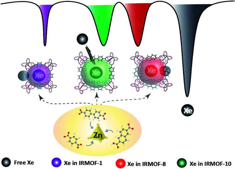
As one of the most important imaging modalities, magnetic resonance imaging (MRI) still faces relatively low sensitivity to monitor low-abundance molecules. A newly developed technology, hyperpolarized 129Xe magnetic resonance imaging (MRI), can boost the signal sensitivity to over 10 000-fold compared with that under conventional MRI conditions, and this technique is referred to as ultrasensitive MRI. However, there are few methods to visualize complex mixtures in this field due to the difficulty in achieving favorable "cages" to capture the signal source, namely, 129Xe atoms. Here, we proposed metal-organic frameworks (MOFs) as tunable nanoporous hosts to provide suitable cavities for xenon. Due to the widely dispersed spectroscopic signals, 129Xe in different MOFs was easily visualized by assigning each chemical shift to a specific color. The results illustrated that the pore size determined the exchange rate, and the geometric structure and elemental composition influenced the local charge experienced by xenon. We confirmed that a complex mixture was first differentiated by specific colors in ultrasensitive MRI. The introduction of MOFs helps to overcome long-standing obstacles in ultrasensitive, multiplexed MRI.


Conflict of interest statement

There are no conflicts to declare.

Figures

Scheme 1
Scheme 1. MOF pore structures are altered to provide diverse microenvironments for 129Xe atom hosting and produce distinguishable MR signals. Multicolor 129Xe MR images are introduced by staining the unique signal in each altered MOF with a specific color.
Fig. 1
Fig. 1. Characterization of the morphology of the MOF nanoparticles. (a) TEM and (b) SEM images of IRMOF-1, IRMOF-8, and IRMOF-10 showed spherical structures and relatively uniform size distributions. Scale bar, 100 nm.
Fig. 2
Fig. 2. Manipulation of the MOF pore structure to produce unique 129Xe MR signals. (a) Frequency-dependent saturation spectra of IRMOF-1, IRMOF-8, and IRMOF-10. To eliminate the influence of solvent, chemical shifts were referenced to the dissolved free 129Xe atom. (b) Coloring ultrasensitive MRI with three types of MOF nanoparticles. Each MOF is saturated at its unique chemical shift, thus enabling discrimination from others in ultrasensitive MRI. (c) Calculating the charge in an edge of an MOF pore. Charge numbers contribute to the differences in chemical shifts. (d) A mixture containing IRMOF-1, IRMOF-8, and IRMOF-10 showed separated signals. (e) MOF nanoparticles provide diverse contrast agents for complex mixture imaging. The MOF mixture showing ultrasensitive MRI in three colors, thus enabling the labeling of three different targets in a single sample.
Fig. 3
Fig. 3. Quantitative measurements of IRMOF nanoparticles. (a) The Hyper-CEST effect of IRMOF-1 at concentrations of 0.05, 0.1, 0.2, 0.5, and 1 mg mL−1. (b) The concentration of IRMOF-1 is linearly related to the Hyper-CEST contrast. (c) The intensity of ultrasensitive 129Xe MRI is enhanced as the concentration increases.

References

    1. Kim D. Park S. Y. Multicolor Fluorescence Photoswitching: Color-Correlated versus Color-Specific Switching. Adv. Opt. Mater. 2018;6:1800678. doi: 10.1002/adom.201800678. - DOI
    1. Mishra K. Stankevych M. Fuenzalida-Werner J. P. Grassmann S. Gujrati V. Huang Y. Klemm U. Buchholz V. R. Ntziachristos V. Stiell A. C. Multiplexed whole-animal imaging with reversibly switchable optoacoustic proteins. Sci. Adv. 2020;6:24. - PMC - PubMed
    1. Wu X. Liu H. Liu J. Haley K. N. Treadway J. A. Larson J. P. Ge N. Peale F. Bruchez M. P. Multicolor Liposome Mixtures for Selective and Selectable Cargo Release. Nano Lett. 2018;18:1331–1336. doi: 10.1021/acs.nanolett.7b05025. - DOI - PubMed
    1. Laviv T. Kim B. B. Chu J. Lam A. J. Lin M. Z. Yasuda R. Simultaneous dual-color fluorescence lifetime imaging with novel red-shifted fluorescent proteins. Nat. Methods. 2016;13:989–992. doi: 10.1038/nmeth.4046. - DOI - PMC - PubMed
    1. Coutu D. L. Kokkaliaris K. D. Kunz L. Schroeder T. Multicolor quantitative confocal imaging cytometry. Nat. Methods. 2018;15:39–46. doi: 10.1038/nmeth.4503. - DOI - PubMed