. 2021 Feb 9;12(12):4494-4502.
doi: 10.1039/d0sc03940c.

Single-step synthesis and interface tuning of core-shell metal-organic framework nanoparticles

Affiliations

Single-step synthesis and interface tuning of core-shell metal-organic framework nanoparticles

Kieran W P Orr et al. Chem Sci. .

Abstract

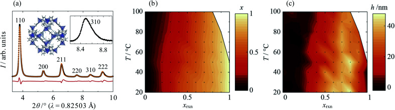
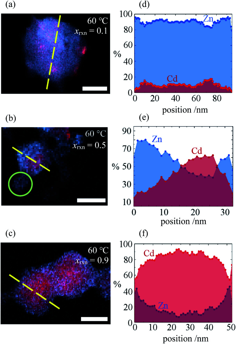
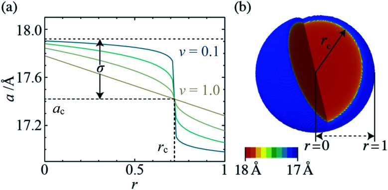
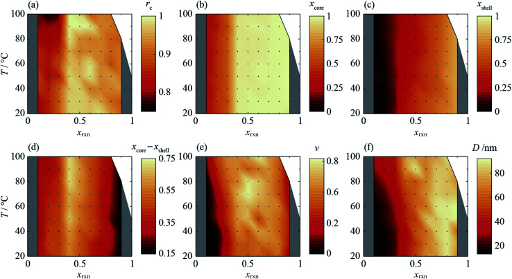
Control over the spatial distribution of components in metal-organic frameworks has potential to unlock improved performance and new behaviour in separations, sensing and catalysis. We report an unprecedented single-step synthesis of multi-component metal-organic framework (MOF) nanoparticles based on the canonical ZIF-8 (Zn) system and its Cd analogue, which form with a core-shell structure whose internal interface can be systematically tuned. We use scanning transmission electron microscopy, X-ray energy dispersive spectroscopy and a new composition gradient model to fit high-resolution X-ray diffraction data to show how core-shell composition and interface characteristics are intricately controlled by synthesis temperature and reaction composition. Particle formation is investigated by in situ X-ray diffraction, which reveals that the spatial distribution of components evolves with time and is determined by the interplay of phase stability, crystallisation kinetics and diffusion. This work opens up new possibilities for the control and characterisation of functionality, component distribution and interfaces in MOF-based materials.


Conflict of interest statement

There are no conflicts to declare.

Figures

Fig. 1
Fig. 1. (a) Single-phase split-peak model fit to the high-resolution synchrotron XRD data, (b) average Cd mole fraction, x, and (c) peak anisotropy, h, as a function of synthesis temperature, T, and reaction Cd mole fraction, xrxn. (a) shows data from synthesis with xrxn = 0.5 at 20 °C (λ = 0.82503 Å). Experimental, calculated and difference data are shown in black, orange and red, respectively, and peaks are labelled with their corresponding Miller indices. Insets show the crystal structure of Zn-ZIF-8 (Zn, C, H and N atoms are shown in dark blue, grey, light grey and light blue, respectively) and an enlarged view of the 310 peak, which exhibits typical peak asymmetry. In (b) and (c), the T-xrxn conditions explored are shown as points and conditions under which phases other than ZIF-8 formed are shown in white.
Fig. 2
Fig. 2. (a–c) STEMEDX images of Zn/Cd ZIF-8 nanoparticles synthesised at T = 60 °C with xrxn = 0.1, 0.5, and 0.9, respectively, and (d–f) line profiles corresponding to dashed lines in (a–c) showing the percentage of Zn and Cd at each pixel. Scale bars = 30 nm; green circle in (b) indicates possible secondary nucleation of a Zn-rich particle.
Fig. 3
Fig. 3. (a) Radial lattice parameter profiles derived using the composition gradient model for ν = 0.1, 0.25, 0.5 and 1.0 (ac = 17.42 Å, rc = 0.72, and σ = 0.5), and (b) 3-D lattice parameter distribution, which corresponds to in situ XRD data for xrxn = 0.5, T = 25 °C, t = 100 s (ac = 17.47 Å, rc = 0.94, σ = 0.52, and ν = 0.13).
Fig. 4
Fig. 4. Radial composition profiles determined by composition gradient model fitting, for xrxn = 0.1, 0.3, 0.5, 0.7 and 0.9 [(a–e), respectively] at T = 20–100 °C. Insets show 3-D composition distributions corresponding to T = 60 °C, sizes scaled to the coherent scattering length, D. Scale bar = 25 nm.
Fig. 5
Fig. 5. Synthesis-structure prediction (SSP) maps for selected structural characteristics of core–shell Zn/Cd ZIF-8 nanoparticles as a function of synthesis temperature, T, and reaction mixture Cd mole fraction, xrxn: (a) nominal interface radius, rc, (b) representative core Cd mole fraction, xcore, calculated from radial composition profiles at r = 0.05, (c) representative shell Cd mole fraction, xshell, calculated at r = 0.95, (d) core–shell composition difference, xcorexshell, (e) interface diffuseness, ν, and (f) coherent scattering length, D.
Fig. 6
Fig. 6. (a) In situ XRD data of mixed Zn/Cd ZIF-8 crystallisation as a function of reaction time, t, for xrxn = 0.5 at T = 25 °C, and (b) the corresponding radial composition profiles. Inset shows the evolution of total diffraction peak intensity, I (beige), interface diffuseness, ν (turquoise), and nominal interface radius, rc (blue).
Fig. 7
Fig. 7. Evolution of core–shell Zn/Cd ZIF-8 particle structure, derived by composition gradient model fits to time-resolved in situ synchrotron XRD data for xrxn = 0.5 at T = 25 °C. Note that particles are not scaled according to size. Stages (1)–(4) represent the nucleation of the Cd-rich core, growth of the Zn-rich shell, increasing interface diffuseness accompanying continued particle growth, and increasing interface diffuseness beyond the end of crystallisation, respectively.

References

    1. Burrows A. D. CrystEngComm. 2011;13:3623. doi: 10.1039/C0CE00568A. - DOI
    1. Abednatanzi S. Gohari Derakhshandeh P. Depauw H. Coudert F.-X. Vrielinck H. Van Der Voort P. Leus K. Chem. Soc. Rev. 2019;48:2535–2565. doi: 10.1039/C8CS00337H. - DOI - PubMed
    1. Wang L. J. Deng H. Furukawa H. Gándara F. Cordova K. E. Peri D. Yaghi O. M. Inorg. Chem. 2014;53:5881–5883. doi: 10.1021/ic500434a. - DOI - PubMed
    1. Deng H. Doonan C. J. Furukawa H. Ferreira R. B. Towne J. Knobler C. B. Wang B. Yaghi O. M. Science. 2010;327:846–850. doi: 10.1126/science.1181761. - DOI - PubMed
    1. Shekhah O. Liu J. Fischer R. A. Wöll C. Chem. Soc. Rev. 2011;40:1081–1106. doi: 10.1039/C0CS00147C. - DOI - PubMed